June 26, 2021

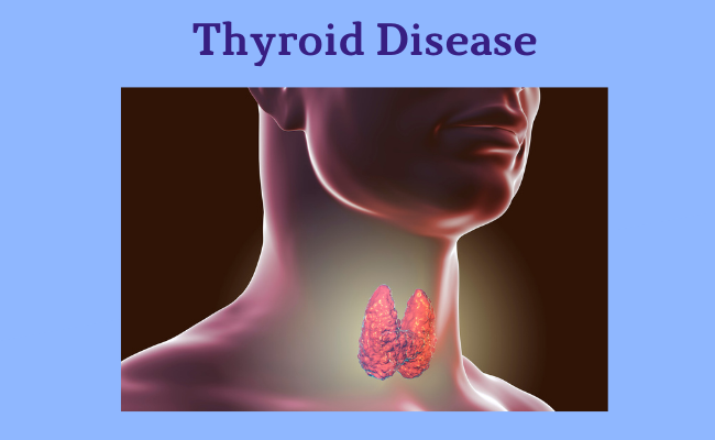
Your thyroid creates and produces hormones that play a role in many different systems throughout your body. When your thyroid makes either too much or too little of these important hormones, it’s called a thyroid disease. 

Thyroid Functions

Your thyroid has an important job to do within your body — releasing and controlling thyroid hormones that control metabolism. Metabolism is a process where the food you take into your body is transformed into energy. This energy is used throughout your entire body to keep many of your body’s systems working correctly.

The thyroid controls your metabolism with a few specific hormones — T4 (thyroxine, contains four iodide atoms) and T3 (triiodothyronine, contains three iodide atoms). These two hormones are created by the thyroid and they tell the body’s cells how much energy to use. When your thyroid works properly, it will maintain the right amount of hormones to keep your metabolism working at the right rate. As the hormones are used, the thyroid creates replacements.

Common Thyroid Problems

The thyroid gland is prone to several very distinct problems, some of which are extremely common. These problems can be broken down into

  1. Those concerning the production of hormone (too much, or too little),
  2. Those due to increased growth of the thyroid, causing compression of important neck structures or simply appearing as a mass in the neck,
  3. The formation of nodules or lumps within the thyroid which are worrisome for the presence of thyroid cancer, and
  4. Those which are cancerous.
  • Goiters: A thyroid goiter is a significantly enlarged thyroid gland that is usually noticed as a bump on the neck. Goiters are often removed for cosmetic reasons as well as to prevent the growth from causes problems such as compressing vital structures of the neck including the trachea and the esophagus. when a goiter grows inward, it may cause a sense of pressure or make it difficult and uncomfortable to breathe and even to swallow. Sometimes thyroid goiters will actually grow into the chest where they can cause trouble as well. Several x-rays will help uncover the cause of any goiter.           
     
  • Thyroid Cancer: Thyroid cancer is a fairly common malignancy, however, the vast majority have excellent long term survival.
     
  • Solitary Thyroid Nodules: There are several characteristics of solitary nodules of the thyroid which make them suspicious for malignancy. Although as many as 50% of the population will have a nodule somewhere in their thyroid, the overwhelming majority of these are benign. Occasionally, thyroid nodules can take on characteristics of malignancy and require either a needle biopsy or surgical excision. 
  • Hyperthyroidism : Hyperthyroidism means too much thyroid hormone. Current methods used for treating a hyperthyroid patient are radioactive iodine, anti-thyroid drugs, or surgery. Each method has advantages and disadvantages and is selected for individual patients. Many times the situation will suggest that all three methods are appropriate, while other circumstances will dictate a single best therapeutic option. Surgery is the least common treatment selected for hyperthyroidism.
     
  • Hypothyroidism: Hypothyroidism means too little thyroid hormone and is a common problem. In fact, hypothyroidism is often present for a number of years before it is recognized and treated. Hypothyroidism can even be associated with pregnancy. Treatment for all types of hypothyroidism is usually straightforward.
     
  • Thyroiditis : Thyroiditis is an inflammatory process ongoing within the thyroid gland. Thyroiditis can present with a number of symptoms such as fever and pain, but it can also present as subtle findings of hypo or hyperthyroidism. There are a number of causes, some more common than others.

Causes of Thyroid Disease

The two main types of thyroid disease are hypothyroidism and hyperthyroidism. Both conditions can be caused by other diseases that impact the way the thyroid gland works.

Conditions that can cause hypothyroidism include:

  • Thyroiditis: This condition is an inflammation (swelling) of the thyroid gland. Thyroiditis can lower the amount of hormones your thyroid produces.
  • Hashimoto’s thyroiditis: A painless disease, Hashimoto’s thyroiditis is an autoimmune condition where the body’s cells attack and damage the thyroid. This is an inherited condition.
  • Postpartum thyroiditis: This condition occurs in 5% to 9% of women after childbirth. It’s usually a temporary condition.
  • Iodine deficiency: Iodine is used by the thyroid to produce hormones. An iodine deficiency is an issue that affects several million people around the world..
  • A non-functioning thyroid gland: Sometimes, the thyroid gland doesn’t work correctly from birth. This affects about 1 in 4,000 newborns. If left untreated, the child could have both physical and mental issues in the future. All newborns are given a screening blood test in the hospital to check their thyroid function.

Conditions that can cause hyperthyroidism include:

  • Graves’ disease: In this condition the entire thyroid gland might be overactive and produce too much hormone. This problem is also called diffuse toxic goiter (enlarged thyroid gland).
  • Nodules: Hyperthyroidism can be caused by nodules that are overactive within the thyroid. A single nodule is called toxic autonomously functioning thyroid nodule, while a gland with several nodules is called a toxic multi-nodular goiter.
  • Thyroiditis: This disorder can be either painful or not felt at all. In thyroiditis, the thyroid releases hormones that were stored there. This can last for a few weeks or months.
  • Excessive iodine: When you have too much iodine (the mineral that is used to make thyroid hormones) in your body, the thyroid makes more thyroid hormones than it needs. Excessive iodine can be found in some medications (amiodarone, a heart medication) and cough syrups.

Common Symptoms of Thyroid Disease

Symptoms of an overactive thyroid (hyperthyroidism) can include:

  • Experiencing anxiety, irritability and nervousness.
  • Trouble sleeping.
  • Losing weight.
  • Enlarged thyroid gland or a goiter.
  • Muscle weakness and tremors.
  • Irregular menstrual periods or Cessation of menstrual cycle.
  • Feeling sensitive to heat.
  • Vision problems or eye irritation.

Symptoms of an underactive thyroid (hypothyroidism) can include:

  • Fatigue or feeling tired.
  • weight gain.
  • Forgetfulness.
  • Episodes of frequent and heavy menstrual periods.
  • Dry and coarse hair.
  • Hoarse voice.
  • Intolerance to cold temperatures.

Diagnosis of Thyroid Disease

Sometimes, thyroid disease can be difficult to diagnose because the symptoms are easily confused with those of other conditions. You may experience similar symptoms when you are pregnant or aging and you would when developing a thyroid disease. Fortunately, there are tests that can help determine if your symptoms are being caused by a thyroid issue. These tests include:

  • Blood tests.
  • Imaging tests.
  • Physical exams.

Blood tests

One of the most definitive ways to diagnose a thyroid problem is through blood tests. Thyroid blood tests are used to tell if your thyroid gland is functioning properly by measuring the amount of thyroid hormones in your blood.

Imaging tests

In many cases, taking a look at the thyroid itself can answer a lot of questions. Your healthcare provider might do an imaging test called a thyroid scan. This allows your provider to look at your thyroid to check for an increased size, shape or growths (nodules).

Your provider could also use an imaging test called an Ultrasound.

Physical exam

Another way to quickly check the thyroid is with a physical exam in your healthcare provider’s office. This is a very simple and painless test where your provider feels your neck for any growths or enlargement of the thyroid.

Treatment of Thyroid Disease

Your healthcare provider’s goal at our clinic in Dubai is to return your thyroid hormone levels to normal. This can be done in a variety of ways and each specific treatment will depend on the cause of your thyroid condition.

If you have high levels of thyroid hormones (hyperthyroidism), treatment options can include:

  • Anti-thyroid drugs (methimazole and propylthioracil): These are medications that stop your thyroid from making hormones.
  • Radioactive iodine: This treatment damages the cells of your thyroid, preventing it from making high levels of thyroid hormones.
  • Beta blockers: These medications don’t change the amount of hormones in your body, but they help control your symptoms.
  • Surgery: A more permanent form of treatment, your healthcare provider may surgically remove your thyroid (thyroidectomy). This will stop it from creating hormones. However, you will need to take thyroid replacement hormones for the rest of your life.

If you have low levels of thyroid hormones (hypothyroidism), the main treatment option is:

  • Thyroid replacement medication: This drug is a synthetic (man-made) way to add thyroid hormones back into your body. One drug that’s commonly used is called levothyroxine. By using a medication, you can control thyroid disease and live a normal life.

Thyroid Removal Surgeries

If your healthcare provider, Dr. Majid Bassuni determines that your thyroid needs to be removed, there are a couple of ways that can be done. Your thyroid may need to be completely removed or just partially. This will depend on the severity of your condition. Also, if your thyroid is very big (enlarged) or has a lot of growths on it, that could prevent you from being eligible for some types of surgery.

The surgery to remove your thyroid is called a thyroidectomy. There are two main ways this surgery can be done:

  • With an incision on the front of your neck.
  • With an incision in your armpit.

Talk to your about all of your treatment options and the best type of surgery for you.

How long does it take to recover from thyroid surgery (thyroidectomy)?

It will take your body a few weeks to recover after your thyroid is surgically removed (thyroidectomy). During this time you should avoid a few things, including:

  • Submerging your incision under water.
  • Lifting an object that’s heavier than 15 pounds.
  • Doing more than light exercise.

This generally lasts for about two weeks. After that, you can return to your normal activities as per your doctors advise.

Should you require additional information or would like to make an appointment with our Consultant General surgeon, Dr. Majid Bassuni ,please call us or e-mail us at info@westminsterclinic.ae

Reference:

  • my.clevelandclinic.org
  • hopkinsmedicine.org
  • Endocrineweb.com
  • Nhs.uk

Disclaimer: All contents on this site are for general information and in no circumstances information be substituted for professional advice from the relevant healthcare professional, Writer does not take responsibility of any damage done by the misuse or use of the information.

June 23, 2021

Examples of functional GI and motility disorders which affect the upper GI tract, from the esophagus to the stomach, include:

Achalasia

Achalasia is a motility disorder in which the esophagus (food tube) empties slowly. The delay results from poor opening of the lower esophageal sphincter (valve) in association with the loss of the normal, orderly muscle activity (peristalsis) that propels foods and liquids along the esophagus into the stomach.

Achalasia results from the nerves in the esophagus and lower esophageal valve being damaged or destroyed. Most cases are idiopathic, meaning the cause is unknown. The origin may possibly be viral or autoimmune, and rarely it may be due to a cancer.

Symptoms of Achalasia

The symptoms of achalasia most often occur during and after a meal. All individuals experience the sensation of solids, and usually liquids, hanging up and passing slowly into the stomach. This may occur several times a week or at every meal.

Effortless regurgitation of bland, undigested food or whitish foam (saliva) is common and may be associated with coughing and choking at night. Some people experience chest pain, and heartburn is common. This heartburn is not related to acid reflux into the esophagus, but rather due to retained acidic food or the fermentation of food in the esophagus.

Treatment of Achalasia

The goal of treating achalasia is to disrupt and open the lower esophageal valve to improve esophageal emptying and relieve symptoms. Unfortunately, no treatment is available to promote the return of peristalsis.

The best treatments for healthy patients are pneumatic dilatation or laparoscopic Heller myotomy. Frail or elderly patients may do well with botulinum toxin (Botox) injections.

Treatment markedly relieves symptoms, but is rarely curative. Overall, the success of both the pneumatic dilation and heller myotomy procedures is 80%-90% and dependent on the skills of the operator. Retreatment may be required and alternative treatments may be needed.

  • Pneumatic dilatation – This procedure involves upper GI endoscopy with the passage of various size balloons to tear the esophagus from within, opening the valve. The procedure is done with conscious sedation and takes about 30 minutes, with a loss of one day of activity. The major complication, esophageal perforation (hole in the esophagus), is rare (less than 5%), but will require major surgery.
  • Heller myotomy – Surgery for achalasia involves cutting the muscle (myotomy) from the outside via small laparoscopic sites on the abdomen. The procedure requires general anesthesia, hospitalization for up to two days, and limited activity for two weeks. The major side effect is cutting the muscle too much, causing bad acid reflux.
  • Botulinum toxin – Botox can be injected into the esophagus and lower esophageal valve, via a needle passed through an upper endoscope. The toxin relaxes the sphincter, and thereby relieves symptoms. In young patients, symptoms relief is generally for only 3-6 months, while older patients may have relief for one year or longer. Adverse events are rare.

Medications may also be tried to relax the lower esophageal sphincter when surgery or pneumatic dilation is not an option, or Botox therapy has failed. Calcium channel blockers and long-acting nitrates are commonly used.

Cyclic Vomiting Syndrome (CVS)

Cyclic vomiting syndrome (CVS) is a disorder with repeated episodes of severe nausea and vomiting that alternate with symptom free periods. It occurs in children and adults.

What are the signs and symptoms of CVS?

The symptom episodes tend to follow the same pattern in each person with CVS over time. There are typically four phases:

  • The first phase is relatively symptom-free. It occurs between vomiting episodes and usually lasts weeks to months.
  • During the second phase the coming on of an episode is felt. There is nausea, but oral medicines may still be taken. This phase lasts minutes to hours.
  • In the third phase there is intense nausea and vomiting, and an inability to eat, drink, or take medicines without vomiting. Other symptoms may include belly pain, hot sweats, cold chills, headache, sensitivity to light and sounds, and diarrhea. The person may be drowsy and withdrawn. This phase lasts from hours to days.
  • In the fourth phase, recovery begins with the settling down of symptoms and ends with going back to a normal diet and a return to the relatively symptom-free period.

Treatment

In general, treatment includes avoiding potential triggering factors, taking medicines to prevent episodes or reduce symptoms, and getting supportive care during episodes.

Triggering factors like stress, anxiety, or certain foods will vary between persons. Try to identify and avoid triggers.

Drug treatments may be divided into short-term treatment of the vomiting episodes and long-term treatment to try to prevent the episodes. In the short term, antiemetic agents can reduce nausea and vomiting. Antianxiety and antimigraine medications may also help. Long term, antidepressant can help prevent nausea and vomiting. Other medicines may also be used as preventive therapies.

Continual vomiting can cause other problems, which need to be treated as well. Examples include loss of fluids (dehydration), electrolyte imbalance, and irritation of the esophagus (food tube).

Dyspepsia

Dyspepsia, also called indigestion, may include symptoms like upper abdominal pain, upper abdominal burning or heat and the stomach feeling full during, or painfully full after, eating.

While dyspeptic symptoms may develop due to diseases such as peptic ulcer or gastritis, the vast majority of people who see doctors for dyspeptic symptoms are ultimately diagnosed as having functional dyspepsia.

Functional dyspepsia (FD) is a heterogeneous disorder; in other words a variety of causes can lead to similar symptoms. Some studies suggest that FD is a chronic condition for many people, with approximately 50% of patients still experiencing symptoms over a 5-year follow-up period.

Treatment Options

Treatment options for functional dyspepsia generally include one or a combination of:

  • Diet
  • Eradication of H. pylori
  • Acid-lowering medications
  • Prokinetic and antiemetic agents
  • Centrally acting therapies

Diet – Most patients with FD have symptoms associated with the ingestion of food. As such, a variety of dietary recommendations are often made, although no clinical trials have formally evaluated specific dietary interventions for the treatment of FD. There are limited data to suggest that dietary fat may induce or exacerbate symptoms and patients often report improvement by eating low-fat meals, and more frequent, smaller meals.

Eradication of H. pylori – Although testing and treating H. pylori infection is recommended and often employed, the eradication of H. pylori infection generally does not improve symptoms outside of those associated with peptic ulcer disease.

Acid-lowering Medications – H2 blockers are the initial agent used by many primary care providers, and are marginally better than placebo at improving upper-center abdominal (epigastric) pain, although they do not improve other dyspeptic symptoms.

If H2 blockers fail to improve symptoms, most doctors then use a proton pump inhibitor (PPI) Short-term risks of this strategy are low, and this may provide relief of symptoms in a small number of people, many of whom may have had silent acid reflux. As a group, PPIs are approximately 10% better than placebo at improving dyspeptic symptoms.

Centrally Acting Therapies – Much of the control of digestive function and sensation is under central nervous system guidance; therapies that target central control mechanisms may have value in relieving symptoms. Tricyclic antidepressants, used in doses lower than required to treat depression, may improve symptoms of dyspepsia in patients who have failed to benefit from treatment with H2 blockers or prokinetics.

Hypnotherapy may improve dyspeptic symptoms in some patients, and a recent study found that hypnotherapy was better than medical therapy and supportive therapy at improving quality of life and symptom scores. Although not well studied, psychological therapies, including cognitive behavioral therapy, may also relieve symptoms of dyspepsia.

Gastroparesis

Gastroparesis is a disorder characterized by symptoms and in which the stomach takes too long to empty its contents. No obstruction or blockage is evident.

In most people diagnosed with gastroparesis, the cause is unknown (idiopathic). Diabetes is the most common known cause of gastroparesis. The condition can also result as a complication from some surgical procedures, from taking certain medications, and from certain other illnesses.

Gastroparesis Treatment

The treatment for gastroparesis in an individual depends on the severity of symptoms. Treatments are aimed at managing symptoms over a long-term.

Treatment approaches may involve one or a combination of:

  • Dietary and lifestyle measures,
  • Medications, and/or
  • procedures that may include surgery, such as
  • Enteral nutrition,
  • Parenteral nutrition,
  • Gastric electrical stimulation (Enterra), or
  • Other surgical procedures

Some people with gastroparesis have mild symptoms that come and go, which can be managed with dietary and lifestyle measures.

GERD

GERD, or gastroesophageal reflux disease, develops when the back-flow (reflux) of stomach contents causes troublesome symptoms and/or complications. Serious health problems can result if it is not treated properly.

The most frequent symptoms of GERD, heartburn and acid regurgitation, are so common that they may not be associated with a disease. Self-diagnosis can lead to mistreatment. Consultation with a physician is essential to proper diagnosis and treatment of GERD.

Treatment for GERD includes:

  • Lifestyle and dietary changes
  • Medication
  • Endoscopic therapy
  • Surgery

Lifestyle and Dietary Changes

Dietary and lifestyle changes are the first step in treating GERD. Certain foods make the reflux worse. Suggestions to help alleviate symptoms include:

  • Lose weight if you are overweight — of all of the lifestyle changes you can make, this one is the most effective.
  • Avoid foods that increase the level of acid in your stomach, including caffeinated beverages.
  • Avoid foods that decrease the pressure in the lower esophagus, such as fatty foods, alcohol and peppermint.
  • Avoid foods that affect peristalsis (the muscle movements in your digestive tract), such as coffee, alcohol and acidic liquids.
  • Avoid foods that slow gastric emptying, including fatty foods.
  • Avoid large meals.
  • Quit smoking.
  • Do not lie down immediately after a meal.
  • Elevate the level of your head when you lie down.

Globus

Globus (Latin for globe or ball) is the sensation of a lump or ball in the throat, typically felt in the throat at the level of the Adam’s apple. It is commonly experienced with an intense emotional experience. The “globus response” is a common human experience. Globus seems equally prevalent in men and women.

Causes of Globus

As with all the functional gastrointestinal disorders, the cause of the globus sensation is unknown. Hypersensitivity of the upper esophagus is suspected. One observer suggested that nervousness leads to a dry mouth, repeated swallowing, and enhanced awareness of the throat.

Distinguishing Globus from Other Conditions

From a medical point of view, it is important to distinguish the globus sensation from dysphagia, or difficulty swallowing. Unlike globus, dysphagia usually indicates a demonstrable cause, and mandates investigation of the esophagus. Heartburn commonly occurs with globus, as it does in people generally. However, the two conditions do not appear to be causally related, and treatment of heartburn does not reliably benefit the globus.

Globus uniquely occurs between meals, and is somewhat relieved by swallowing something, often a glass of water. On the other hand, dysphagia occurs during the swallowing of food or sometimes liquids. It gives the sensation that something is stuck in the gullet – often below the throat. An attempt to swallow in this situation seems to make things worse.

“Red Flag” Signs and Symptoms that may indicate a More Serious Disorder

  • Neck or throat pain
  • Bleeding from the mouth or throat
  • Weight loss
  • Pain or difficulty on swallowing
  • Muscle weakness
  • Mass in the throat or mouth confirmed by a doctor’s examination
  • Progressive worsening of the symptom

Treatment of Globus

By itself, globus seldom indicates a structural disease, that is a disease where damage to the body can be demonstrated. Nevertheless, any accompanying symptom must be investigated as indicated. There is no specific treatment for globus beyond the assurance that it isn’t the sign of a serious disease.

Should you require additional information or would like to make an appointment with our Consultant General surgeon, Dr. Majid Bassuni ,please call us or e-mail us at info@westminsterclinic.ae

Reference:

  • Iffgd.org – International Foundation for Gastrointestinal Disorders
  • hopkinsmedicine.org
  • National Institute of Diabetes and Digestive and Kidney Diseases
  • gastro.org: American Gastroenterological Association

Disclaimer: All contents on this site are for general information and in no circumstances information be substituted for professional advice from the relevant healthcare professional, Writer does not take responsibility of any damage done by the misuse or use of the information.

June 21, 2021

Dry needling is a type of alternative medicine treatment wherein hollow-core hypodermic needles or solid filiform needles are used to give relief to muscle pain. In addition, it also helps in movement impairments. In this treatment, needles are inserted through the skin into the affected areas of the muscle. It is also known as myofascial trigger point dry needling, intramuscular manual therapy, or Intramuscular Stimulation  (IMS). Dry needling is a part of modern Western medicine principles and supported by research.

How is it performed?

 A practitioner who is usually a Physical Therapist certified in dry needling, will insert numerous filiform short and fine needles into your skin during dry needling. This technique is “dry” which means that no fluid will be injected in the body.

Physical therapists wear gloves and appropriate personal protective equipment (PPE) when dry needling, consistent with Standard Precautions, Guide to Infection Prevention for Outpatient Settings. The sterile needles are disposed of in a medical sharps collector. The technique can be only carried on upon obtaining the patient signed consent form. In cases when physical therapists use dry needling, it is typically 1 technique that’s part of a larger treatment plan.

Why dry Needling?

Physical therapists use dry needling to release trigger points to relieve pain or improve range of motion. This can help speed up the patient’s return to active rehabilitation.

The needles will be placed in “Myofascial trigger points” which are hyperirritable spots in skeletal muscle that are associated with a hypersensitive palpable nodule in a taut band. The Trigger points can be the cause of pain, range of movement restriction and muscle weakness. Trigger point dry needling can be carried out at superficial or deep tissue level.

Benefits of Dry Needling

Dry needling is primarily used to treat myofascial trigger points.

“Trigger points” refers to the tight bands of skeletal muscles situated within a very large muscle group. The points are tender to touch and when you touch a particular “trigger point”, it may cause pain to some other parts of your body. These “trigger points” are nothing but local contractures in a muscle fiber that restrict your range of motion or disrupt function. Dry needling helps in increasing blood flow, reducing referred and local pain, and decreasing overall tightness or banding.

Among the countless health issues that dry needling can help resolve, the most pivotal ones are as follows:

  • Joint problems
  • Disc problems
  • Tendinitis
  • Migraine and tension-type headaches
  • Jaw and mouth problems (such as temporomandibular joint disorders or TMD)
  • Whiplash
  • Repetitive motion disorders (like carpal tunnel syndrome)
  • Pelvic pain
  • Night cramps
  • Phantom pain
  • Post-herpetic neuralgia (pain left behind by shingles)
  • Low back pain/tension
  • Neck pain
  • Tennis elbow
  • Migraines
  • Spinal dysfunction
  • Sciatica

Should you require additional information or would like to make an appointment with our Physiotherapist ,please call us or e-mail us at info@westminsterclinic.ae

Disclaimer: All contents on this site are for general information and in no circumstances information be substituted for professional advice from the relevant healthcare professional, Writer does not take responsibility of any damage done by the misuse or use of the information.

June 20, 2021

The gallbladder is a small pouch-like organ found underneath the liver. Its main purpose is to store and concentrate bile. Bile is a liquid produced by the liver to help digest fats. It’s passed from the liver into the gallbladder through a series of channels known as bile ducts.

Gallstones are hard, pebble-like pieces of material, usually made of cholesterol or bilirubin, that develop in the gallbladder. When gallstones block your bile ducts, they can cause sudden pain, which means you need medical attention right away.

Causes of Gallstones

Gallstones are thought to develop because of an imbalance in the chemical make-up of bile inside the gallbladder. In most cases the levels of cholesterol in bile become too high and the excess cholesterol forms into stones.

  • A greater amount of cholesterol or bilirubin in bile.
  • Hormones or medications that result in decreased emptying of the gallbladder.
  • Obesity.
  • Inactive lifestyle.
  • Being female.
  • Being over 40 years old.
  • Diabetes.
  • Liver disease.
  • Family history of gallstones.

Symptoms of Gallstones

Gallstones often have no symptoms. But if a gallstone becomes trapped in an opening (duct) inside the gallbladder, it can trigger a sudden, intense pain in your tummy that usually lasts between 1 and 5 hours. This type of abdominal pain is known as biliary colic.

Some people with gallstones can also develop complications, such as inflammation of the gallbladder (cholecystitis).

This can cause:

  • Pain in the upper mid abdomen or upper right abdomen.
  • Associated pain in the right shoulder.
  • Chest pain.
  • Nausea and vomiting.
  • Repeated similar episodes.
  • Yellowing of the skin and eyes (jaundice)
  • A high temperature

Diagnosis

Your Doctor at Westminster Ortho Med Clinic, Dubai may use Perform:

  • Taking Medical history
  • Physical examination
  • Lab and imaging tests to diagnose and find gallstones.
  • Blood tests can show signs of infection or inflammation of the bile ducts, gallbladder, pancreas, or liver.

Treatment

Silent stones are not treated and should be left alone. Most people who have gallstones are in this category.If you have symptoms, such as pain, you will probably need to be treated.

The most common treatment for gallstones is to remove the gallbladder surgically. Removal of the gallbladder is called a cholecystectomy. In the majority of cases (90%), this surgery can be performed laparoscopically, a minimally invasive technique that results in less post-operative pain and a faster recovery than conventional cholecystectomy.

Gallstones that are found in the bile ducts may need to be removed even if there are no symptoms. This procedure is commonly done with the endoscopic retrograde cholangiopancreatography (ERCP) procedure.

Laparoscopic Cholecystectomy

Laparoscopic cholecystectomy is known as a minimally invasive procedure because it uses several small incisions instead of one large one. A laparoscope is a narrow tube with a camera. This surgical tool is inserted through one incision. The camera allows your doctor to see your gallbladder on a TV screen. Your gallbladder is then removed through another small incision.

Endoscopic Retrograde Cholangiopancreatography (ERCP) Procedure

An endoscopic retrograde cholangiopancreatography (ERCP) procedure is another minimally invasive option for removing gallstones. The endoscope is a tube-like tool. Your doctor will need to move it throughout your digestive system during the treatment. The path the endoscope travels is:

  • In the mouth.
  • Down the throat.
  • Through the stomach.
  • Into the duodenum (beginning of the small intestine) where the common bile duct empties its bile.

Once there, the endoscope is used to remove any blockages in the bile duct.

Non-surgical treatments for gallstones

In most cases where gallstones need treatment, your healthcare provider will use a minimally invasive technique to remove the stone.

There are medications that can be used to dissolve the stones. However, as minimally invasive methods have advanced, these drugs haven’t been used as often. Dissolving medications can take months—or possibly even years—to get rid of the gallstones.

By contrast, a procedure resolves the issue quickly. Using these medications to treat gallstones could be an option if you are unable to have surgery because of another condition. Talk to your doctor about all treatment options and which one is the best fit for you.

Foods to avoid if you have had gallstones in the past

Many gallstones are formed from cholesterol. Though you cannot prevent gallstones, you can try and limit the amount of fatty foods in your diet. Several tips for limiting cholesterol in your diet include:

  • Eating less meat.
  • Adding fish.
  • Limiting the amount of fried foods.
  • Adding more whole grains.
  • Choosing low-fat dairy products (cheese, milk).
  • Adding fresh vegetables and fruit.

Should you require additional information or would like to make an appointment with our Consultant General surgeon, Dr. Majid Bassuni ,please call us or e-mail us at info@westminsterclinic.ae

Reference:

  • American Gastroenterological Association
  • nhs.uk
  • National Institute of Diabetes and Digestive and Kidney Diseases
  • Clevelandclinic.org

Disclaimer: All contents on this site are for general information and in no circumstances information be substituted for professional advice from the relevant healthcare professional, Writer does not take responsibility of any damage done by the misuse or use of the information.

June 16, 2021

A fistula is an abnormal connection or passageway that connects two organs or vessels that do not usually connect. They can develop anywhere between an intestine and the skin, between the vagina and the rectum, and other places. The most common location for a fistula is around the anus.

Fistulas may occur in many parts of the body. They can form between:

  • An artery and vein
  • Bile ducts and the surface of the skin (from gallbladder surgery)
  • The cervix and vagina
  • The neck and throat
  • The space inside the skull and nasal sinus
  • The bowel and vagina
  • The colon and surface of the body, causing feces to exit through an opening other than the anus
  • The stomach and surface of the skin
  • The uterus and peritoneal cavity (the space between the walls of the abdomen and internal organs)
  • An artery and vein in the lungs (results in blood not picking up enough oxygen in the lungs)
  • The navel and gut

TYPES OF FISTULAS:

ANAL FISTULAS/PERIANAL FISTULAS.

Abnormal connection between the epithelialized surface of the anal canal and the perianal skin.

  • Anorectal Fistula occurs between the anal canal and the skin around the anal opening.
  • Rectovaginal or Anovaginal Fistula occurs when a hole develops between the rectum or anus and the vagina.
  • Colovaginal Fistula occurs between the colon and the vagina.

URINARY TRACT FISTULAS.

Abnormal openings within a urinary tract organ or an abnormal connection between a urinary tract organ and another organ.

  • Vesicouterine fistula occurs between the bladder and the uterus.
  • Vesicovaginal fistula is where a hole develops between the bladder and the vagina.
  • Urethrovaginal fistula is between the urethra and the vagina.

OTHER TYPES.

  • Enteroenteral fistula occurs between two parts of the intestine.
  • Enterocutaneous or Colocutaneous fistula occurs between the small intestine and the skin or the colon and the skin respectively.

CAUSES OF FISTULA

Crohn’s disease and diverticular disease are well known to cause fistula formation. In addition, those who are undergoing radiation therapy are at greater risk for a variety of fistulas. 

Most anal fistulas develop after an anal abscess. One can develop fistula if the abscess does not heal properly after the pus has drained away.

Less common causes of anal fistulas include:

  • Crohn’s disease – a long-term condition in which the digestive system becomes inflamed
  • Diverticulitis – infection of the small pouches that can stick out of the side of the large intestine (colon)
  • Hidradenitis suppurativa – a long-term skin condition that causes abscesses and scarring
  • Infection with tuberculosis (TB) or HIV
  • A complication of surgery near the anus

WHAT ARE THE SYMPTOMS OF FISTULAS?

Depending on the type of fistula, here are some of the signs and symptoms:

  • Constant urine leakage from the vagina
  • Irritation in the external female genital organs
  • Frequent urinary tract infections (UTIs)
  • Leakage of gas and/or feces into the vagina
  • Fluid drainage from the vagina
  • Nausea
  • Vomiting
  • Diarrhea
  • Abdominal pain

Patients can have different symptoms depending on which parts of the body are connected by the fistula.

Fistula between the small and large intestine

  • Diarrhea
  • Passage of undigested food

Fistula between the intestine and the bladder

  • Urinary tract infection
  • Burning with urination
  • Cloudy urine or blood in the urine

Fistula between the intestine and the vagina

  • Passage of gas or stool through the vagina

Fistula from the intestine to the skin

  • Can initially present as a painful bump or boil
  • Skin abscess that is open and draining fluid or stool

If you have one or more of these symptoms, be sure to promptly discuss them with a your Doctor, Dr. Majid Bassuni, at our clinic in Dubai.

FISTULA TREATMENT OPTIONS

Following the diagnosis of a fistula, the medical professional will decide the best plan of treatment based on its location, size, and condition. One treatment path may simply be controlling symptoms with a catheter. More severe fistula repair may require surgery.

MANAGEMENT

Non-invasive Treatment

  • Fibrin glue. A specific medicinal adhesive used to seal fistulas.
  • Plug. This is usually a collagen matrix used to fill the fistula.
  • Catheters. Used to drain fistulas, catheters are usually employed on small fistulas to manage infection.

SURGICAL TREATMENT

  • Transabdominal surgery. The fistula is accessed through an abdominal wall incision.
  • Laparoscopic surgery. This is a minimally invasive surgery that involves a tiny incision and the use of cameras and small tools to repair the fistula.

PHARMACEUTICAL TREATMENT

Antibiotics or other medication may also be used to treat any infection associated with the fistula. Yet there is no pharmaceutical solution to eradicate fistulas at this time.

While fistulas pose a serious threat to your body, high treatment success should encourage you or someone dealing with fistulas to seek immediate help.

Ask Your Doctor

  • What are my options for fistula removal?
  • What preparations will I need to make before my fistula removal surgery?
  • What are the potential complications from surgery?
  • What kind of restrictions will I have after my surgery?
  • How long will it take me to recover from fistula removal surgery?
  • How will the surgery affect my diet and bowel movements?
  • If I require an ileostomy, how do I care for my ostomy pouch and keep it clean?
  • What supplies will I need at home?
  • Will I need additional surgeries?

Should you require additional information or would like to make an appointment with our Consultant General/Colorectal Surgeon, Dr. Majid Bassuni ,please call us or e-mail us at info@westminsterclinic.ae

Reference:

  • crohnscolitisfoundation.org
  • nhs.uk
  • American Society of Colon and Rectal Surgeons (ASCRS)
  • National Association for Continence (nafc.org)

Disclaimer: All contents on this site are for general information and in no circumstances information be substituted for professional advice from the relevant healthcare professional, Writer does not take responsibility of any damage done by the misuse or use of the information.

May 29, 2021

Scoliosis is an abnormal curvature of the spine (backbone). There is a natural, forward-and-backward curve to the spine. With scoliosis, the spine rotates and develops a side-to-side curve.

Most cases of scoliosis are mild and don’t need treatment. In adults, the degree of the spinal curve may or may not determine treatment. Treatment is geared towards relieving symptoms, and not necessarily fixing the curve. The goal is always to decrease pain and improve function.

Types of Scoliosis

Idiopathic scoliosis is scoliosis without a known cause. In as many as 80% of cases, doctors don’t find the exact reason for a curved spine.

Congenital scoliosis begins as a baby’s back develops before birth. Problems with the tiny bones in the back, called vertebrae, can cause the spine to curve. The vertebrae may be incomplete or fail to divide properly. Doctors may spot this rare condition when the child is born. Or they may not find it until the teen years.

Neuromuscular scoliosis is caused by a disorder like spina bifidacerebral palsy, or a spinal cord injury. These conditions sometimes damage your muscles so they don’t support your spine correctly. That can cause your back to curve.

Degenerative scoliosis affects adults. It usually develops in the lower back as the disks and joints of the spine begin to wear out as you age.

Causes and Risk Factors

Some kinds of scoliosis have clear causes. Doctors divide those curves into two categories — structural and nonstructural.

In nonstructural scoliosis, the spine works normally but looks curved. This happens for a number of reasons, such as having one leg that’s longer than the other, muscle spasms, and inflammations like appendicitis. When these problems are treated, the scoliosis often goes away.

In structural scoliosis, the curve of the spine is rigid and can’t be reversed.

Causes include:

For idiopathic scoliosis, family history and genetics can be risk factors. If you or one of your children has this condition, make sure your other kids are checked regularly.

Signs of Scoliosis

Oftentimes scoliosis is first suspected when someone notices something slightly off and comments. Some examples could include:

  • Clothes fit awkwardly or hang unevenly.  Person with scoliosis might notice that a shirt or blouse appears uneven, which could be cause for further investigation.
  • Sideways curvature observed while in bathing suit or changing. For instance, a parent could first notice the sideways curvature in an adolescent’s back while at the pool or beach.

Some of the more common symptoms present in moderate or severe scoliosis could include:

  • Changes with walking. When the spine abnormally twists and bends sideways enough, it can cause the hips to be out of alignment, which changes a person’s gait or how they walk. The extra compensating that a person does to maintain balance for the uneven hips and legs can cause the muscles to tire sooner. A person might also notice that one hand brushes against a hip while walking but the other does not.
  • Reduced range of motion. The deformity from spinal twisting can increase rigidity, which reduces the spine’s flexibility for bending.
  • Trouble breathing. If the spine rotates enough, the rib cage can twist and tighten the space available for the lungs. Bone might push against the lungs and make breathing more difficult.
  • Cardiovascular problems. Similarly, if the rib cage twists enough, reduced spacing for the heart can hamper its ability to pump blood.
  • Pain. If curvature becomes severe enough, back muscles could become more prone to painful spasms. Local inflammation may develop around the strained muscles, which can also lead to pain. It is possible for the intervertebral discs and facet joints to start to degenerate due to higher loads.
  • Lower self-esteem. This symptom is commonly overlooked or minimized by outside observers, but it can be a significant factor for people who have a noticeable spinal deformity. Especially for adolescents who want to fit in with their friends, it can be stressful and depressing to look different, have clothes fit unevenly, or wear a noticeable back brace that may be uncomfortable or limit activity.

Scoliosis Treatment

For mild scoliosis, you may not need treatment. Instead, your doctor at Westminster clinic, Dubai might watch you and take X-rays once in a while to see if it’s getting worse. Some children grow out of scoliosis.

If you or your child need treatment, your doctor might suggest:

  • Braces. In kids who are still growing, wearing a brace around your torso can stop the curve from getting worse. They’re usually made of plastic. Many kids wear them 24 hours a day. You can’t see them under clothes, and they don’t stop you from doing everyday activities.

Choosing Observation vs. Bracing

About 90% of idiopathic scoliosis cases are mild and only require observation. Typically, observation is done by a doctor every 4 to 6 months until the adolescent has reached full skeletal maturity.

Brace Options

There are two general treatment options for scoliosis bracing:

  • Full-time bracing. These braces are designed to be worn 16 to 23 hours a day. The goal is to wear them all the time with exceptions for bathing, skin care, and exercising.
  • Nighttime bracing. These braces use hyper-corrective forces (which put the body out of normal balance and cannot realistically be applied while a person is standing and/or performing daily activities) and are to be worn at least 8 hours a night.
  • Spinal fusion surgery. In this operation, your doctor puts pieces of bone or a similar material between bones in your spine. They use hardware to hold the bones in place until they grow together, or fuse. The surgery can lessen the curve in your spine as well as keep it from getting worse.
  • Spine and rib-based growing operation. This is done to correct more serious scoliosis in children who are still growing. The doctor attaches rods to your spine or ribs with hardware. As you grow, the doctor adjusts the length of the rods.

Should you require additional information or would like to make an appointment with our Consultant Orthopedic Surgeon Dr. Ishrat Khan Pain Consultant Dr. Tarek Sultan OR Physical Therapists,  Anil Daniel,  OR Hadel Radwan  please call us or e-mail us at info@westminsterclinic.ae

Reference:

  • clevelandclinic.org
  • fda.gov
  • orthoinfo.aaos.org – American Academy of Orthopedic Surgeons
  • Mayoclinic.org
  • nhs.uk

Disclaimer: All contents on this site are for general information and in no circumstances information be substituted for professional advice from the relevant healthcare professional, Writer does not take responsibility of any damage done by the misuse or use of the information.

May 16, 2021

Metabolic syndrome is a cluster of conditions that occur together, increasing your risk of heart disease, stroke and type 2 diabetes. These conditions include increased blood pressure, high blood sugar, excess body fat around the waist, and abnormal cholesterol or triglyceride levels.

Having just one of these conditions doesn’t mean you have metabolic syndrome. But it does mean you have a greater risk of serious disease. The term “metabolic” refers to the biochemical processes involved in the body’s normal functioning. Risk factors are traits, conditions, or habits that increase your chance of developing a disease.

Risk factors

The following factors increase your chances of having metabolic syndrome:

  • Age. Your risk of metabolic syndrome increases with age.
  • Ethnicity. In the United States, Hispanics — especially Hispanic women — appear to be at the greatest risk of developing metabolic syndrome. The reasons for this are not entirely clear. Some racial and ethnic groups in the United States are at higher risk for metabolic syndrome than others. Mexican Americans have the highest rate of metabolic syndrome, followed by whites and blacks.

Other groups at increased risk for metabolic syndrome include:

  • People who have a personal history of diabetes
  • People who have a sibling or parent who has diabetes
  • Women when compared with men
  • Women who have a personal history of polycystic ovarian syndrome (a tendency to develop cysts on the ovaries)
  • Obesity. Carrying too much weight, especially in your abdomen, increases your risk of metabolic syndrome.
  • Diabetes. You’re more likely to have metabolic syndrome if you had diabetes during pregnancy (gestational diabetes) or if you have a family history of type 2 diabetes.
  • Other diseases. Your risk of metabolic syndrome is higher if you’ve ever had nonalcoholic fatty liver disease, polycystic ovary syndrome or sleep apnea.

Symptoms of metabolic syndrome

Metabolic syndrome may be diagnosed if you have 3 or more of the following:

  • Being very overweight or having too much fat around your waist
  • High triglyceride levels (fat in the blood) and low levels of HDL (the “good” cholesterol) in your blood, which can lead to atherosclerosis (where arteries become clogged with fatty substances such as cholesterol)
  • High blood pressure that’s consistently 140/90mmHg or higher
  • An inability to control blood sugar levels (insulin resistance)

Causes- Metabolic Syndrome

Metabolic syndrome has several causes that act together. You can control some of the causes, such as overweight and obesity, an inactive lifestyle, and insulin resistance.

You can’t control other factors that may play a role in causing metabolic syndrome, such as growing older. Your risk for metabolic syndrome increases with age.

You also can’t control genetics (ethnicity and family history), which may play a role in causing the condition. For example, genetics can increase your risk for insulin resistance, which can lead to metabolic syndrome.

Researchers continue to study conditions that may play a role in metabolic syndrome, such as:

  • A fatty liver (excess triglycerides and other fats in the liver)
  • Polycystic ovarian syndrome (a tendency to develop cysts on the ovaries)
  • Gallstones
  • Breathing problems during sleep (such as sleep apnea)

Diagnosis

Your doctor will diagnose metabolic syndrome based on the results of a physical exam and blood tests. You must have at least three of the five metabolic risk factors to be diagnosed with metabolic syndrome.

Treatment

Heart-healthy lifestyle changes are the first line of treatment for metabolic syndrome. If heart-healthy lifestyle changes aren’t enough, your doctor may prescribe medicines. Medicines are used to treat and control risk factors, such as high blood pressure, high triglycerides, low HDL (“good”) cholesterol, and high blood sugar.

Goals of Treatment

The major goal of treating metabolic syndrome is to reduce the risk of ischemic heart disease. Treatment is directed first at lowering LDL cholesterol and high blood pressure and managing diabetes (if these conditions are present).

The second goal of treatment is to prevent the onset of type 2 diabetes, if it hasn’t already developed. Long-term complications of diabetes often include heart and kidney disease, vision loss, and foot or leg amputation. If diabetes is present, the goal of treatment is to reduce your risk for heart disease by controlling all of your risk factors.

Heart-Healthy Lifestyle Changes 

Heart-healthy lifestyle changes include heart-healthy eatingaiming for a healthy weightmanaging stressphysical activity, and quitting smoking.  

Medicines

Sometimes lifestyle changes aren’t enough to control your risk factors for metabolic syndrome.  For example, you may need statin medications to control or lower your cholesterol. By lowering your blood cholesterol level, you can decrease your chance of having a heart attack or stroke. Doctors usually prescribe statins for people who have:

  • Diabetes
  • Heart disease or had a prior stroke
  • High LDL cholesterol levels

Dr. Saima Salahuddin in our clinic in Dubai may discuss beginning statin treatment with those who have an elevated risk for developing heart disease or having a stroke.

Your doctor also may prescribe other medications to:

  • Decrease your chance of having a heart attack.
  • Lower your blood pressure.
  • Prevent blood clots, which can lead to heart attack or stroke.
  • Reduce your heart’s workload and relieve symptoms of coronary heart disease.

Take all medicines regularly, as your doctor prescribes. Don’t change the amount of your medicine or skip a dose unless your doctor tells you to. You should still follow a heart-healthy lifestyle, even if you take medicines to treat your risk factors for metabolic syndrome. 

Prevention

A lifelong commitment to a healthy lifestyle may prevent the conditions that cause metabolic syndrome. A healthy lifestyle includes:

  • Getting at least 30 minutes of physical activity most days
  • Eating plenty of vegetables, fruits, lean protein and whole grains
  • Limiting saturated fat and salt in your diet
  • Maintaining a healthy weight
  • Not smoking

Should you require additional information or would like to make an appointment with our Consultant Family Medicine, Dr. Saima Salahuddin ,please call us or e-mail us at info@westminsterclinic.ae

Reference:

  • NIH – National Lung, Heart and Blood Institute
  • nhs.uk
  • Mayoclinic.org
  • American Heart Association

Disclaimer: All contents on this site are for general information and in no circumstances information be substituted for professional advice from the relevant healthcare professional, Writer does not take responsibility of any damage done by the misuse or use of the information.

April 28, 2021

Whiplash is also known as neck sprain or neck strain. This common type of neck injury happens when the neck jolts backward or forward, sharply and suddenly. Whiplash due to a motor vehicle collision or another injury can strain your muscles or damage soft tissues in your neck. 

What causes a whiplash?

Whiplash typically occurs when your head is forcefully and quickly thrown backward and then forward. This motion can injure bones in the spine, disks between the bones, ligaments, muscles, nerves and other tissues of the neck.

A whiplash injury may result from:

  • Auto accidents. Rear-end collisions are a major cause of whiplash.
  • Physical abuse or assault. Whiplash can occur if you are punched or shaken. It’s one of the injuries seen in shaken baby syndrome.
  • Contact sports. Football tackles and other sports-related collisions can sometimes cause whiplash.

Symptoms

Signs and symptoms of whiplash usually develop within days of the injury, and may include:

  • Neck pain and stiffness
  • Worsening of pain with neck movement
  • Loss of range of motion in the neck
  • Headaches, most often starting at the base of the skull
  • Tenderness or pain in the shoulder, upper back or arms
  • Tingling or numbness in the arms
  • Fatigue
  • Dizziness

Some people also have:

  • Blurred vision
  • Ringing in the ears (tinnitus)
  • Sleep disturbances
  • Irritability
  • Difficulty concentrating
  • Memory problems
  • Depression

Diagnosis

Along with a complete medical history and physical exam, tests for whiplash may include the following. Many whiplash injuries include damage to soft tissue that can’t be seen on X-rays:

  • X-ray. Electromagnetic energy beams produce images of internal tissues, bones, and organs onto film.
  • Magnetic resonance imaging (MRI). Large magnets and a computer make detailed images of organs and soft tissue structures in your body.
  • Computed tomography (CT) scan. X-rays and computer technology make horizontal, or axial, images (often called slices) of your body. A CT scan shows detailed images of any part of your body, including your bones, muscles, fat, and organs. CT scans are more detailed than general X-rays.

Treatment

Your healthcare provider at Westminster Ortho Med Clinic, DHCC, Dubai will determine specific treatment for whiplash, based on:

  • Your age, overall health, and medical history
  • Extent of your injury
  • Your tolerance for specific medicines, procedures, or therapies
  • Expectations for the course of your injury
  • Your opinion or preference

Pain management

Your doctor may recommend one or more of the following treatments to lessen pain:

  • Rest. Rest may be helpful for a day or two after your injury, but too much bed rest may delay recovery.
  • Heat or cold. Either heat or cold applied to the neck for 15 minutes every three hours or so can help you feel better.
  • Over-the-counter pain medications. Over-the-counter pain relievers, such as acetaminophen (Tylenol, others) and ibuprofen (Advil, Motrin IB, others), often can control mild to moderate whiplash pain.
  • Prescription medications. People with more-severe pain may be given certain antidepressant drugs that have been shown to relieve nerve pain.
  • Muscle relaxants. Short-term use of these drugs may be recommended to loosen tight muscles and soothe pain. The medicine also can make you feel sleepy. It may be used to help restore normal sleep if pain prevents you from getting a good night’s rest.
  • Injections. An injection of lidocaine (Xylocaine) — a numbing medicine — into painful muscle areas may be used to decrease pain so that you can do physical therapy.

Exercise

Your doctor will likely prescribe a series of stretching and movement exercises for you to do at home. These exercises can help restore range of motion in your neck and get you back to your normal activities. Applying moist heat to the painful area or taking a warm shower may be recommended before exercise.

Exercises may include:

  • Rotating your neck in both directions
  • Tilting your head side to side
  • Bending your neck toward your chest
  • Rolling your shoulders

Physical therapy

If you have ongoing whiplash pain or need assistance with range-of-motion exercises, your doctor may recommend that you see a physical therapist. Physical therapy can help you feel better and may prevent further injury. Your physical therapist will guide you through exercises to strengthen your muscles, improve posture and restore normal movement.

In some cases, transcutaneous electrical nerve stimulation (TENS) may be used. TENS applies a mild electric current to the skin. Limited research suggests this treatment may temporarily ease neck pain and improve muscle strength.

The number of physical therapy sessions needed will vary from person to person. Your physical therapist can also create a personalized exercise routine that you can do at home.

Foam collars

Soft foam cervical collars were once commonly used for whiplash injuries to hold the neck and head still. However, studies have shown that keeping the neck still for long periods of time can decrease muscle strength and interfere with recovery.

Still, use of a collar to limit movement may help reduce pain soon after your injury, and may help you sleep at night. Recommendations for using a collar vary though. Some experts suggest limiting use to no more than 72 hours, while others say it may be worn up to three hours a day for a few weeks. Your doctor can instruct you on how to properly use the collar, and for how long.

Alternative medicine

Nontraditional therapies have been tried to treat whiplash pain, but research about how well they work is limited. Some include:

  • Acupuncture. Acupuncture involves inserting ultrafine needles through specific areas on your skin. It may offer some relief from neck pain.
  • Chiropractic care. A chiropractor performs joint manipulation techniques. There is some evidence that chiropractic care may provide pain relief when paired with exercise or physical therapy. Manipulation of the spine may cause minor problems, such as numbness or dizziness, and rarely damage to spinal tissues.
  • Massage. Neck massage may provide short-term relief of neck pain from whiplash injury.
  • Mind-body therapies. Exercises that incorporate gentle movements and a focus on breathing and mindfulness, such as tai chi, qi gong and yoga, may help ease pain and stiffness.

Should you require additional information or would like to make an appointment with our Consultant Orthopedic Surgeon Dr. Ishrat Khan Pain Consultant Dr. Tarek Sultan OR Physical Therapists,  Anil Daniel,  OR Hadel Radwan  please call us or e-mail us at info@westminsterclinic.ae

Reference:

  • clevelandclinic.org
  • Johns Hopkins Medicine
  • orthoinfo.aaos.org – American Academy of Orthopedic Surgeons
  • Mayoclinic.org

Disclaimer: All contents on this site are for general information and in no circumstances information be substituted for professional advice from the relevant healthcare professional, Writer does not take responsibility of any damage done by the misuse or use of the information.

April 27, 2021

Spinal stenosis (or narrowing) is a common condition that occurs when the small spinal canal, which contains the nerve roots and spinal cord, becomes compressed. This causes a “pinching” of the spinal cord and/or nerve roots, which leads to pain, cramping, weakness or numbness. Depending on where the narrowing takes place, you may feel these symptoms in the lower back and legs, neck, shoulder or arms.

Causes

Some people are born with a small spinal canal. This is called “congenital stenosis”. However, spinal narrowing is most often due to age-related changes that take place over time. This is called “acquired spinal stenosis.”

The risk of developing spinal stenosis increases if:

  • You were born with a narrow spinal canal
  • You are female
  • You are 50 years old or older
  • You’ve had a previous injury or surgery of the spine

Some medical conditions can cause spinal stenosis. These include:

  • Osteoarthritis and bony spurs that form as we age
  • Inflammatory spondyloarthritis (e.g., ankylosing spondylitis)
  • Spinal fractures and injuries
  • Spinal tumors
  • Paget’s Disease

Symptoms:

Back pain. People with spinal stenosis may or may not have back pain, depending on the degree of arthritis that has developed.

Burning pain in buttocks or legs (sciatica). Pressure on spinal nerves can result in pain in the areas that the nerves supply. The pain may be described as an ache or a burning feeling. It typically starts in the area of the buttocks and radiates down the leg. As it progresses, it can result in pain in the foot.

Numbness or tingling in buttocks or legs. As pressure on the nerve increases, numbness and tingling often accompany the burning pain. Although not all patients will have both burning pain and numbness and tingling.

Weakness in the legs or “foot drop.” Once the pressure reaches a critical level, weakness can occur in one or both legs. Some patients will have a foot-drop, or the feeling that their foot slaps on the ground while walking.

Less pain with leaning forward or sitting. Studies of the lumbar spine show that leaning forward can actually increase the space available for the nerves. Many patients may note relief when leaning forward and especially with sitting. Pain is usually made worse by standing up straight and walking. Some patients note that they can ride a stationary bike or walk leaning on a shopping cart. Walking more than 1 or 2 blocks, however, may bring on severe sciatica or weakness.

Diagnosis:

Medical History and Physical Examination

After discussing your symptoms and medical history, your doctor at Westminster Ortho Med Clinic, DHCC, Dubai will examine your back. This will include looking at your back and pushing on different areas to see if it hurts. Your doctor may have you bend forward, backward, and side-to-side to look for limitations or pain.

Imaging Tests

Other tests which may help your doctor, Dr. Ishrat Khan OR Dr. Tarek Sultan to confirm your diagnosis include:

X-rays. Although they only visualize bones, X-rays can help determine if you have spinal stenosis. X-rays will show aging changes, like loss of disk height or bone spurs.

X-rays taken while you lean forward and backward can show “instability” in your joints. X-rays can also show too much mobility. This is called spondylolisthesis.

Magnetic resonance imaging (MRI). This study can create better images of soft tissues, such as muscles, disks, nerves, and the spinal cord.

Additional tests. Computed tomography (CT) scans can create cross-section images of your spine. Your doctor may also order a myelogram. In this procedure, dye is injected into the spine to make the nerves show up more clearly. It can help your doctor determine whether the nerves are being compressed.

Treatment Options:

Nonsurgical Treatment

Nonsurgical treatment options focus on restoring function and relieving pain. Although nonsurgical methods do not improve the narrowing of the spinal canal, many people report that these treatments do help relieve symptoms.

Exercise
Regular exercise can help you build and maintain strength in the muscles of your arms and upper legs (the hip adductors and abductors, quadriceps and hamstrings). This will improve your balance, ability to walk, bend and move about, as well as control pain. A physical therapist can show you which exercises are right for you.

Physical therapy. Stretching exercises, massage, and lumbar and abdominal strengthening often help manage symptoms.

A physical therapist, Ms. Hadel Radwan can teach you exercises that may help:

  • Build up your strength and endurance
  • Maintain the flexibility and stability of your spine
  • Improve your balance

Lumbar traction. Although it may be helpful in some patients, traction has very limited results. There is no scientific evidence of its effectiveness.

Anti-inflammatory medications. Because stenosis pain is caused by pressure on a spinal nerve, reducing inflammation (swelling) around the nerve may relieve pain. Non-steroidal anti-inflammatory drugs (NSAIDs) initially provide pain relief. When used over the course of 5-10 days, they can also have an anti-inflammatory effect.

Steroid injections. Steroid injections around the nerves or in the “epidural space” can decrease swelling, as well as pain. They also reduce numbness, but not weakness, in the legs. Patients should receive no more than three injections a year.

Acupuncture. Acupuncture can be helpful in treating some of the pain for less severe cases of lumbar stenosis. Although it can be very safe, long-term success with this treatment has not been proven scientifically.

Surgical Treatment

Surgery for lumbar spinal stenosis is generally reserved for patients who have poor quality of life due to pain and weakness. Patients may complain of difficulty walking for extended periods of time. This is often the reason that patients consider surgery.

There are two main surgical options to treat lumbar spinal stenosis: laminectomy and spinal fusion. Both options can result in excellent pain relief. Be sure to discuss the advantages and disadvantages of both with your doctor.

Laminectomy. This procedure involves removing the bone, bone spurs, and ligaments that are compressing the nerves. This procedure may also be called a “decompression.” Laminectomy can be performed as open surgery, in which your doctor uses a single, larger incision to access your spine. The procedure can also be done using a minimally invasive method, where several smaller incisions are made. Your doctor will discuss the right option for you.

Spinal fusion. If arthritis has progressed to spinal instability, a combination of decompression and stabilization or spinal fusion may be recommended.

Rehabilitation. After surgery, you may stay in the hospital for a short time, depending on your health and the procedure performed. Healthy patients who undergo just decompression may go home the same or next day, and may return to normal activities after only a few weeks. Fusion generally adds 2 to 3 days to the hospital stay.

Your surgeon may give you a brace or corset to wear for comfort. He or she will likely encourage you to begin walking as soon as possible. Most patients only need physical therapy to strengthen their backs.

Your physical therapist may show you exercises to help you build and maintain strength, endurance, and flexibility for spinal stability. Some of these exercises will help strengthen your abdominal muscles, which help support your back. Your physical therapist will create an individualized program, taking into consideration your health and history.

Surgical risks. There are minor risks associated with every surgical procedure. These include bleeding, infection, blood clots, and reaction to anesthesia. These risks are usually very low.

Surgical outcomes. Overall, the results of laminectomy with or without spinal fusion for lumbar stenosis are good to excellent in the majority of patients. Patients tend to see more improvement of leg pain than back pain. Most patients are able to resume a normal lifestyle after a period of recovery from surgery.

Living with Spinal Stenosis:

Spinal stenosis can lead to the slow but steady loss of strength in the legs. The severe pain caused by this condition can be quite disabling, even if you have no muscle weakness, since it greatly affects your ability to work and enjoy life. The natural course of the disease is one of slow progression over time. There is no cure for this condition but there are steps you can take to feel better. For example:

  • Get moving. Regular exercise is very important, so do it often – at least three times a week for about 30 minutes. Start slowly with flexion-based (forward-bending) exercises. As you begin to feel stronger, add walking or swimming to your plan.
  • Modify your activity. Don’t do anything that can trigger or worsen pain and disability such as lifting heavy objects or walking long distances.
  • Talk to your physician about pain medications, as well as alternative therapies such as acupuncture or massage that can ease pain.
  • Explore non-surgical options first except in rare cases when pain, weakness and numbness comes on quickly.

Should you require additional information or would like to make an appointment with our Consultant Orthopedic Surgeon Dr. Ishrat Khan Pain Consultant Dr. Tarek Sultan OR Physical Therapists,  Anil Daniel,  OR Hadel Radwan  please call us or e-mail us at info@westminsterclinic.ae

Reference:

  • clevelandclinic.org
  • Rhematology.org- American College of Rheumatology
  • orthoinfo.aaos.org – American Academy of Orthopedic Surgeons
  • Mayoclinic.org

Disclaimer: All contents on this site are for general information and in no circumstances information be substituted for professional advice from the relevant healthcare professional, Writer does not take responsibility of any damage done by the misuse or use of the information.

April 26, 2021

Rheumatoid arthritis is a chronic inflammatory disorder that can affect more than just your joints. In some people, the condition can damage a wide variety of body systems, including the skin, eyes, lungs, heart and blood vessels. An autoimmune disorder, rheumatoid arthritis occurs when your immune system mistakenly attacks your own body’s tissues.

32106553 – rheumatoid arthritis (ra) is an inflammatory type of arthritis that usually affects knees. rheumatoid arthritis of the knee the auto immune disease. the body’s immune system mistakenly attacks healthy tissue.

Causes

Rheumatoid arthritis occurs when your immune system attacks the synovium — the lining of the membranes that surround your joints.

The resulting inflammation thickens the synovium, which can eventually destroy the cartilage and bone within the joint. The tendons and ligaments that hold the joint together weaken and stretch. Gradually, the joint loses its shape and alignment.

The cause of RA is not known. There is evidence that autoimmune conditions run in families. For instance, certain genes that you are born with may make you more likely to get RA.

Signs and Symptoms

With Rheumatoid Arthritis, there are times when symptoms get worse, known as flares, and times when symptoms get better, known as remission.

Signs and symptoms of Rheumatoid Arthritis include:

  • Pain or aching in more than one joint
  • Stiffness in more than one joint
  • Tenderness and swelling in more than one joint
  • The same symptoms on both sides of the body (such as in both hands or both knees)
  • Weight loss
  • Fever
  • Fatigue or tiredness
  • Weakness

Diagnosis

Rheumatoid Arthritis is diagnosed at our clinic in Dubai by examining blood test results, physical examination of the joints and reviewing x-ray or ultrasound images. There is no one test to diagnose RA. Blood tests are run to look for antibodies in the blood that can been seen in RA. Antibodies are small proteins in the bloodstream that help fight against foreign substances called antigens. Sometimes these antibodies are found in people without RA. This is called a false positive result. Blood tests are also run to look for high levels of inflammation. The symptoms of RA can be very mild making the diagnosis more difficult. Some viral infections can cause symptoms that can be mistaken for RA. A rheumatologist is a physician with the skill and knowledge to reach a correct diagnosis of RA and to recommend a treatment plan.

Abnormal blood tests commonly seen in RA include:

  • Anemia (a low red blood cell count)
  • Rheumatoid factor (an antibody, or blood protein, found in about 80% of patients with RA in time, but in as few as 30% at the start of arthritis)
  • Antibodies to cyclic citrullinated peptides (pieces of proteins), or anti-CCP for short (found in 60 – 70% of patients with RA)
  • Elevated erythrocyte sedimentation rate (a blood test that, in most patients with RA, confirms the amount of inflammation in the joints)

X-rays can help in detecting RA, but may be normal in early arthritis. Even if normal, initial X-rays may be useful later to show if the disease is progressing. MRI and ultrasound scanning can be done to help confirm or judge the severity of RA.

RA is a chronic arthritis. Generally the symptoms will need to be present for more than three months to consider this diagnosis. However there are patients who are diagnosed sooner.

Treatment

There is no cure for rheumatoid arthritis. But clinical studies indicate that remission of symptoms is more likely when treatment begins early with medications known as disease-modifying anti-rheumatic drugs (DMARDs).

Medications

The types of medications recommended by your doctor will depend on the severity of your symptoms and how long you’ve had rheumatoid arthritis.

  • NSAIDs. Non-steroidal anti-inflammatory drugs (NSAIDs) can relieve pain and reduce inflammation.
  • Steroids. Corticosteroid medications, reduce inflammation and pain and slow joint damage. Side effects may include thinning of bones, weight gain and diabetes. Doctors often prescribe a corticosteroid to relieve acute symptoms, with the goal of gradually tapering off the medication.
  • Disease-modifying anti-rheumatic drugs (DMARDs). These drugs can slow the progression of rheumatoid arthritis and save the joints and other tissues from permanent damage.
  • Biologic agents. Also known as biologic response modifiers, this newer class of DMARDs includes abatacept.

These drugs can target parts of the immune system that trigger inflammation that causes joint and tissue damage. These types of drugs also increase the risk of infections

Therapy

Your doctor at Westminster Ortho Med clinic, Dubai may refer you to a physical therapist who can teach you exercises to help keep your joints flexible. The therapist may also suggest new ways to do daily tasks, which will be easier on your joints. For example, you may want to pick up an object using your forearms.

Assistive devices can make it easier to avoid stressing your painful joints. For instance, a kitchen knife equipped with a hand grip helps protect your finger and wrist joints. Certain tools, such as buttonhooks, can make it easier to get dressed. Catalogs and medical supply stores are good places to look for ideas.

Surgery

If medications fail to prevent or slow joint damage, you and your doctor may consider surgery to repair damaged joints.

Rheumatoid arthritis surgery may involve one or more of the following procedures:

  • Synovectomy. Surgery to remove the inflamed lining of the joint (synovium) can be performed on knees, elbows, wrists, fingers and hips.
  • Tendon repair. Inflammation and joint damage may cause tendons around your joint to loosen or rupture. Your surgeon may be able to repair the tendons around your joint.
  • Joint fusion. Surgically fusing a joint may be recommended to stabilize or realign a joint and for pain relief when a joint replacement isn’t an option.
  • Total joint replacement. During joint replacement surgery, your surgeon removes the damaged parts of your joint and inserts a prosthesis made of metal and plastic.

Living with rheumatoid arthritis

It is important to be physically active most of the time, but to sometimes scale back activities when the disease flares. In general, rest is helpful when a joint is inflamed, or when you feel tired. At these times, do gentle range-of-motion exercises, such as stretching. This will keep the joint flexible.

When you feel better, RA patients are encouraged to do low-impact aerobic exercises, such as walking, and exercises to boost muscle strength. This will improve your overall health and lower the pressure on your joints. A physical therapist, Hadel Radwan OR Anil Daniel at westminster Clinic, Dubai can help you find which types of activities are best for you, and at what level or pace you should do them.

Finding that you have a chronic illness is a life-changing event. It can cause worry and sometimes feelings of isolation or depression. Thanks to greatly improved treatments, these feelings tend to decrease with time as energy improves, and pain and stiffness decrease. Discuss these normal feelings with your health care providers. They can provide helpful information and resources.

Rheumatoid Arthritis Diet

Your healthcare provider or dietitian may recommend an anti-inflammatory diet to help with your symptoms. This type of diet includes foods that have lots of omega-3 fatty acids.

Foods high in omega-3 fatty acids include:

  • Fatty fish like salmon, tuna, herring, and mackerel
  • Chia seeds
  • Flax seeds
  • Walnuts

Antioxidants, such as vitamins A, C, and E, and selenium, may also help reduce inflammation. Foods high in antioxidants include:

  • Berries, such as blueberries, cranberries, goji berries, and strawberries
  • Dark chocolate
  • Spinach
  • Kidney beans
  • Pecans
  • Artichokes

Eating lots of fiber is also important. According to the researchers, fiber may help reduce inflammatory responses which may decrease C-reactive protein levels. Choose whole grain foods, fresh vegetables, and fresh fruit. Strawberries may be particularly beneficial.

Foods containing flavonoids can also help to counter inflammation in the body. They include:

  • Soy products, such as tofu and miso
  • Berries
  • Green tea
  • Broccoli
  • Grapes

What you don’t eat is just as important as what you do eat. Make sure to avoid trigger foods. These include processed carbohydrates and saturated or trans-fats. Avoiding trigger foods and choosing the right foods in trying to follow an anti-inflammatory diet may help you manage your Rheumatoid Arthritis.

Should you require additional information or would like to make an appointment with our Consultant Orthopedic Surgeon Dr. Ishrat Khan Pain Consultant Dr. Tarek Sultan OR Physical Therapists,  Anil Daniel,  OR Hadel Radwan  please call us or e-mail us at info@westminsterclinic.ae

Reference:

  • clevelandclinic.org
  • Center for diseases control and prevention (CDC)
  • yourphysio.org.uk
  • Mayoclinic.org
  • American college of rheumatology

Disclaimer: All contents on this site are for general information and in no circumstances information be substituted for professional advice from the relevant healthcare professional, Writer does not take responsibility of any damage done by the misuse or use of the information.

Call Now Button