February 27, 2021

Most sports injuries are due to either trauma or overuse of muscles or joints. The majority are caused by minor trauma involving muscles, ligaments, tendons, or bones, including:

  • Contusions (bruises)
  • Sprains
  • Strains
  • Fractures
  • Dislocations

Some examples of strains are:

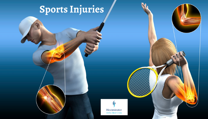
  • Tennis elbow (lateral epicondylitis). Lateral epicondylitis, also known as tennis elbow, is characterized by pain in the backside of the elbow and forearm, along the thumb side when the arm is alongside the body with the thumb turned away. The pain is caused by damage to the tendons that bend the wrist backward away from the palm.
  • Golfer’s or baseball elbow (medial epicondylitis). Medial epicondylitis, also known as golfer’s elbow, is characterized by pain from the elbow to the wrist on the palm side of the forearm. The pain is caused by damage to the tendons that bend the wrist toward the palm.
  • Lumbar strain. A lumbar strain is an injury to the lower back, which results in damaged tendons and muscles that spasm and feel sore. Trauma of great force can injure the tendons and muscles in the lower back. Pushing and pulling sports, such as weight lifting or football, can lead to a lumbar strain. In addition, sports that need sudden twisting of the lower back, such as basketball, baseball, and golf can lead to this injury.
  • Jumper’s knee. Jumper’s knee, also known as patellar tendonitis, is a condition characterized by inflammation of the patellar tendon, which connects the kneecap to shin bone (tibia). The condition may be caused by overuse of the knee joint, such as frequent jumping on hard surfaces.
  • Runner’s knee. Runner’s knee, also known as patellofemoral stress syndrome, is when the patella, or kneecap, does not move well in the groove of the femur (thigh bone). Runner’s knee may be caused by a structural defect, or a certain way of walking or running.

What to do if you have an injury

If you’ve injured yourself, you may have immediate pain, tenderness, swelling, bruising, and restricted movement or stiffness in the affected area. Sometimes, these symptoms may only be noticeable several hours after exercising or playing sports.

Stop exercising if you feel pain, regardless of whether your injury happened suddenly or you’ve had the pain for a while. Continuing to exercise while injured may cause further damage and slow your recovery.

If you have a minor injury, you do not usually need to see a doctor and can look after yourself at home. However, you may want to visit a GP, for advice if your symptoms do not get better over time.

Treating Sports Injury

You can usually treat common minor injuries yourself by:

  • Resting the affected part of the body for the first 48 to 72 hours to prevent further damage
  • Regularly applying an ice pack to the affected area during the first 48 to 72 hours to reduce swelling
  • Using over the counter pain relievers to relieve pain

If your symptoms are severe or do not improve within a few days or weeks, your family physician may be able to refer you for specialist treatment and support, such as physiotherapy OR Sport physiotherapy expert.

Prevention of Sports Injury

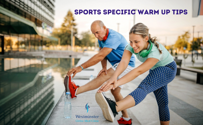
Sports Injuries are common and can often be attributed to missing out important steps such as the ‘warm-up’ and the ‘cool down’.  Failing to warm up first and cool down afterwards may increase the chances of suffering an injury, which could potentially lead to problems like osteoarthritis in later life. The joints are the areas that take a huge amount of pressure, along with the muscles, so making sure we take care of those areas is crucial.  Warm-up really acts to reduce the stress on those areas, reduce the potential for injury and keep us safe while we’re exercising.

If you are taking up a new sport or a regular attendee to the gym to help you avoid injury here are some top tips from the Chartered Society of Physiotherapy on warming up properly.

Rehabilitation for sports injuries

A rehabilitation program for sports injuries is designed to meet the needs of the individual patient, depending on the type and severity of the injury. Active involvement of the patient and family is vital to the success of the program.

The goal of rehabilitation after an amputation is to help the patient return to the highest level of function and independence possible, while improving the overall quality of life–physically, emotionally, and socially.

In order to help reach these goals, sports injury rehabilitation programs may include the following:

  • Activity restrictions
  • Physical or occupational therapy
  • Exercise programs to stretch and strengthen the area
  • Conditioning exercises to help prevent further injury
  • Heat or cold applications and whirlpool treatments
  • Applications of braces, splints, or casts to immobilize the area
  • Use of crutches or wheelchairs
  • Pain management techniques
  • Patient and family education

Should you require additional information or would like to make an appointment with our Consultant Orthopedic Surgeon Dr. Ishrat Khan OR Physical Therapists,  Anil Daniel,  OR Hadel Radwan  please call us or e-mail us at info@westminsterclinic.ae

Reference:

  • oxfordhealth.nhs.uk
  • Hopkinsmedicine.org
  • yourphysio.org.uk
  • Mayoclinic.org

Disclaimer: All contents on this site are for general information and in no circumstances information be substituted for professional advice from the relevant healthcare professional, Writer does not take responsibility of any damage done by the misuse or use of the information.

February 22, 2021

The colon (or large Intestine) can develop small pouches, which bulge outward through the colon’s muscular wall (see diagram below). These pouches develop in areas where blood vessels are entering the muscular wall, which is a weaker spot that allows these pouches to form.

Each pouch is called a diverticulum. Pouches are called diverticula. The condition of having diverticula is called diverticulosis.

When the pouches become infected or inflamed, the condition is called diverticulitis. This occurs in 10 to 25 in every 100 people with diverticulosis. Diverticulosis and diverticulitis are also known as diverticular disease.

What causes diverticular disease?

If your stools become small and hard instead of soft and bulky, your colon has to squeeze hard to push the stool forward. These contractions create high pressure in the muscular wall of your colon and, if this continues over the years, then these pouches develop at the weak areas in the wall of
your colon. This is one of the reasons why diverticula are more likely to develop in the left side of the colon.


There is no exact cause of diverticular disease but the following have been shown to increase your risk of developing the disease – increasing age, obesity, high BMI (body mass index), and people on low fiber diets.
Diverticular disease is common in developed or industrialised countries – particularly the United States, England, and Australia – where low fibre diets are common. The disease is rare in the countries of Asia and Africa, where people eat high-fibre vegetable diets.


Fibre is the part of fruits, vegetables, and grains that the body cannot digest. Some fibre dissolves easily in water (soluble fibre). It takes on a soft, jelly-like texture in the intestines. Some fibre passes almost unchanged through the intestines (insoluble fibre). Both kinds of fibre help make stools soft and easy to pass. Fibre also prevents constipation.

What are the symptoms?


Diverticulosis

Most people with diverticulosis do not have any discomfort or symptoms. However, possible symptoms may include mild cramps, bloating, and constipation. Other diseases such as irritable bowel syndrome (IBS) and stomach ulcers cause similar problems, so these symptoms do not
always mean a person has diverticulosis. You should visit your doctor if you have these symptoms.


Diverticulitis

The most common symptom of diverticulitis is abdominal pain. The most common sign is tenderness around the left side of the lower abdomen. If infection is the cause, fever, nausea (feeling sick), vomiting, chills, cramping, and constipation may occur as well. How bad your
symptoms are depends on the extent of the infection and complications.

What are the complications?


Diverticulitis can lead to bleeding, infections, perforations (small holes), tears, or blockages. These complications always need treatment to prevent them from progressing and causing serious illness.


• Bleeding

Bleeding from diverticula is a rare complication. When diverticula bleed, blood may appear in the toilet or in your stool. Bleeding can be severe, but it may stop by itself and not need treatment. Bleeding diverticula are caused by a small blood vessel in a diverticulum that weakens and finally bursts. If you have bleeding from your rectum, you should see your doctor. If the bleeding does not stop, surgery may be needed.


• Abscess, perforation, and peritonitis

The infection causing diverticulitis often clears up after a few days of treatment with antibiotics. If the condition gets worse, an abscess may form in your colon. An abscess is an infected area with pus that may cause swelling and destroy tissue. Sometimes the infected diverticula may
develop small holes called perforations. These perforations allow pus to leak out of the colon into your abdominal area. If the abscess is small and remains in your colon, it may clear up after treatment with antibiotics. If not, your doctor may need to drain it.


• Fistula

A fistula is an abnormal connection of tissue between two organs or between an organ and the skin. When damaged tissues come into contact with one another during infection, they sometimes stick together. If they heal that way, a fistula forms. When diverticulitis related infections spread outside the colon, the colon tissue may stick to other nearby tissues. The
organs usually involved are the bladder, small intestine, and skin.

The most common type of fistula happens between the bladder and the colon. It affects men more than women. This type of fistula can result in a severe, long-lasting infection of the urinary tract. The problem can be corrected with surgery to remove the fistula and the affected part of your colon.


• Intestinal obstruction

The scarring caused by infection may cause part or total blockage of your colon. When this happens, your colon is unable to move bowel contents normally. When the obstruction totally blocks your colon, emergency surgery is needed. Partial blockage is not an emergency, so the surgery to correct it can be planned.

How does your doctor diagnose diverticular disease?

To diagnose diverticular disease, your doctor will ask about your medical history, perform a physical examination, and may perform one or more tests. As most people do not have symptoms, diverticulosis is often found through tests ordered for another condition.
When taking a medical history, your doctor may ask you about your bowel habits, symptoms, pain, diet, and medications. The physical examination usually involves a digital rectal examination. To perform this test, your doctor inserts a gloved, lubricated finger into your rectum to check for any
tenderness, blockages, or blood. Your doctor may check your stools for signs of bleeding and test your blood for signs of infection.


Your doctor will order further tests which may include a:
colonoscopy or flexible sigmoidoscopy: where a tube with a camera is passed into your colon through your back passage, to view the inner lining of your whole colon. A sigmoidoscopy is a similar procedure that views mostly the left side of your colon. Medication can be given to
make the procedure comfortable and you will be given something before your procedure to make sure you completely empty your bowels.
CT virtual colonoscopy is an x-ray-based test although, as with the colonoscopy, you will
need to completely clean your bowel with bowel preparation the day before.

What is the treatment for diverticula disease?


A high-fibre diet and occasionally, mild painkillers will help relieve symptoms in most cases. Sometimes an attack of diverticulitis is serious enough to need a hospital stay and surgery.


Diverticulosis: Increasing the amount of fibre in your diet may reduce symptoms of diverticulosis and prevent complications such as diverticulitis. Fibre keeps stools soft and lowers pressure inside the colon so that bowel content can move through easily. Twenty to 35 grams of fibre is recommended daily.

Your doctor may also recommend taking a fibre product . Foods such as nuts, popcorn hulls, and sunflower, pumpkin, caraway, and sesame seeds should be avoided. The seeds in tomatoes, courgettes, cucumbers,
strawberries, and raspberries, as well as poppy seeds, are generally considered harmless. People differ in the amounts and types of foods they can eat. Decisions about your diet should be made based on what works best for you. Keeping a diary may help identify individual items in your diet. If cramps, bloating, and constipation are problems, your doctor may prescribe a short course of pain medication. However, many medications can cause constipation.
You should take regular exercise, lose weight if you are overweight or obese, and stop smoking. Although studies do not show that these changes will definitely help all patients, changes to a person’s lifestyle will bring additional wider benefits in general health and wellbeing.

Diverticulitis: Treatment for diverticulitis focuses on clearing up the infection and inflammation, resting your colon, and preventing or minimizing complications. An attack of diverticulitis without complications
may respond to antibiotics within a few days, if treated early enough.
To help your colon rest, your doctor may recommend bed rest and a liquid diet, along with pain relief.

When is surgery necessary?


If your attacks are severe or happen often, your doctor may advise surgery. The surgeon removes the affected part of your colon and joins the remaining sections. This type of surgery is called colon resection, and aims to keep attacks from coming back and prevents complications. Your
doctor may also recommend surgery for complications of a fistula or intestinal obstruction.

Points to remember


• Diverticulosis occurs when small pouches, called diverticula, bulge outward through weak spots in your colon (large intestine).
• The pouches form when pressure inside your colon builds, usually because of constipation.
• Most people with diverticulosis never have any discomfort or symptoms.
• The most likely cause of diverticulosis is a low fibre diet because it increases the likelihood of constipation and pressure inside your colon.
• For most people with diverticulosis, eating a high-fibre diet is the only treatment needed.
• You can increase your fibre intake by eating foods such as wholegrain breads and cereals; fruit like apples and peaches; vegetables like kidney beans and lima beans.
• Diverticulitis occurs when the pouches become infected or inflamed and cause pain and tenderness around the left side of your lower abdomen.

References:

  • NHS www.nhs.uk/live-well/eat-well/
  • British Dietetic Association (BDA) www.bda.uk.com/food-health/food-facts.html

Disclaimer: All contents on this site are for general information and in no circumstances information be substituted for professional advice from the relevant healthcare professional, Writer does not take responsibility of any damage done by the misuse or use of the information.


January 31, 2021

Warming up before exercise can increase blood flow to muscles and other soft tissues gradually, which is an important factor in avoiding damage to ligaments, muscles, joints and tendons. A general warm up should include five to ten minutes of light cardiovascular activity, such as fast walking or slow jogging, to increase your heart rate gradually, promote blood flow to your muscles and supply them with more oxygen.

To properly prepare your muscles and increase your mobility, physiotherapists suggest any warm up routine should include time spent on performing the exercise you’re about to undertake at a slow pace.

Typical examples include a slow jog for running, a gentle cycle for a spin class or a slow swim before building up speed in the pool. Follow this with some sports-specific movements relevant to the activity you’re about to perform. Try service motions for tennis, catching practice for cricket or side stepping for football.

Tennis

  • Shoulder, hip and knee injuries are common amongst tennis players. Mimic the movements of tennis, including high-steps, arm circles, serves and raquet swings in a controlled manner.
  • Lunge walk – Move forwards with your legs in a long, exaggerated stride pattern.
  • Keep your upper body straight but move your arms in time with your legs.
  • Buttock kicks – At slow jogging pace, bring your heels up to meet your bottom.
  • Jumping high and from side to side.
  • Rotating your torso from side to side.


Football

Knees and ankle injuries are common amongst footballers. In addition to your cardiovascular warm up, aim to build the following moves into your preparation.

  • Lunge walk – Move forwards with your legs in a long, exaggerated stride pattern. Keep your upper body straight but move your arms in time with your legs.
  • Side stepping / sideways running – This movement will help to stretch the hips and inner thighs.
  • Running backwards – Carefully running backwards can help prepare the quad and calf muscles before you begin to play.
  • Buttock kicks – At slow jogging pace, bring your heels up to meet your bottom.
  • Chest hugs – Bring your knees up and hug them towards your chest


Running/Jogging:

Knee, ankle and foot injuries are common amongst runners. In addition to your cardiovascular warm up, try the following.

  • Lunge walk – Move forwards with your legs in a long, exaggerated stride pattern. Keep your upper body straight but move your arms in time with your legs.
  • Buttock kicks – At slow jogging pace, bring your heels up to meet your bottom.
  • Controlled leg swings – Standing on your left leg swing your right leg backwards and forwards in a controlled manner. Swap legs.
  • Fast feet – Stand with your feet together, and then lift them quickly off the floor one after the other as if running over hot coals.


Cycling


Knee and lower back problems are common amongst cyclists. Before you get on your bike, jog on the spot, jump from side to side, lunge forwards and back and then try the following move while lying on the floor…
Bring your knees up towards your chest and perform slow paced pedaling movements with your legs.
Once you’re on your bike begin pedaling at a moderate pace, increasing speed and intensity until you reach your desired pace.


Cooling down after exercise


Taking the time to cool down after exercise is important and helps:
• Your heart and breathing rates return towards resting levels gradually
• Prevent you feeling faint or dizzy
• Remove waste products from your muscles which can result in muscle soreness
• Prepare your body for your next exercise session.


Cool down by gradually slowing the pace and effort you’re putting into your chosen form of exercise. Keep going at this reduced pace for five to ten minutes. Another option is to slow jog, brisk walk, or march on the spot for the same length of time.
The activities suggested above should not cause any harm. If you do experience pain or discomfort, stop immediately and speak to a health professional, such as a physiotherapist or your family doctor.

Should you require additional information or would like to make an appointment with our Physical Therapists,  Anil Daniel,  OR Hadel Radwan  please call us or e-mail us at info@westminsterclinic.ae

Reference:

  • Charted Society of physiotherapy – https://www.csp.org.uk/
  • yourphysio.org.uk

Disclaimer: All contents on this site are for general information and in no circumstances information be substituted for professional advice from the relevant healthcare professional, Writer does not take responsibility of any damage done by the misuse or use of the information.

January 30, 2021

Natural anti-inflammatories are foods that you can eat to lower your odds of having inflammation. If you have a condition that causes inflammation, it may help to change your eating habits.

Any mainstream nutrition expert would encourage you to eat anti-inflammatory foods. They include lots of fruits and vegetables, whole grains, plant-based proteins (like beans and nuts), fatty fish, and fresh herbs and spices.

Fruits and veggies: Go for variety and lots of color. Research shows that vitamin K-rich leafy greens like spinach and kale reduce inflammation, as do broccoli and cabbage. So does the substance that gives fruits like cherries, raspberries, and blackberries their color, olives and olive oil.

Whole grains: Oatmeal, brown rice, whole-wheat bread, and other unrefined grains tend to be high in fiber, and fiber also may help with inflammation.

Beans: They’re high in fiber, plus they’re loaded with antioxidants and other anti-inflammatory substances.

Nuts: They have a healthy kind of fat that helps stop inflammation. (Olive oil and avocados are also good sources.) Stick to just a handful of nuts a day, or otherwise the fat and calories will add up.

Fish: Put it on your plate at least twice a week. Salmon, tuna, and sardines all have plenty of omega-3 fatty acids, which fight inflammation.

Herbs and spices: They add antioxidants (along with flavor) to your food. Turmeric, found in curry powder, does this with a strong substance called curcumin. And garlic curbs your body’s ability to make things that boost inflammation.

Who can it help?

An anti-inflammatory diet may serve as a complementary therapy for many conditions that become worse with chronic inflammation.

The following conditions involve inflammation:

  • Rheumatoid arthritis
  • Psoriasis
  • Asthma
  • Eosinophilic esophagitis
  • Crohn’s disease
  • Colitis
  • Inflammatory bowel disease
  • Lupus
  • Hashimoto’s thyroiditis
  • Metabolic syndrome

Metabolic syndrome refers to a collection of conditions that tend to occur together, including type 2 diabetes, obesity, high blood pressure, and cardiovascular disease.

Foods that cause inflammation

Try to avoid or limit these foods as much as possible:

  • Refined carbohydrates, such as white bread and pastries
  • French fries and other fried foods
  • Soda and other sugar-sweetened beverages
  • Red meat (burgers, steaks) and processed meat (hot dogs, sausage)
  • Margarine, shortening, and lard

Risks of Chronic Inflammation

Inflammation happens naturally in your body. Inflammation protects against toxins, infection, and injury, but when it happens too often it can trigger diseases. Experts link long-term (chronic) inflammation to:

  • Cancer
  • Heart disease 
  • Diabetes
  • Alzheimer’s disease
  • Depression 

Anti-inflammatory diet tips

It can be challenging to transition to a new way of eating, but the following tips may help:

  • Pick up a variety of fruits, vegetables, and healthful snacks during the weekly shop.
  • Gradually replace fast food meals with healthful, homemade lunches.
  • Replace soda and other sugary beverages with still or sparkling mineral water.

Other tips include:

  • Talking to a healthcare professional about supplements, such as cod liver oil or a multivitamin.
  • Incorporating 30 minutes of moderate exercise into the daily routine.
  • Practicing good sleep hygiene, as poor sleep can worsen inflammation.

Takeaway

An anti-inflammatory diet may help reduce inflammation and improve symptoms of some common health conditions, such as rheumatoid arthritis.

There is no single anti-inflammatory diet, but a diet that includes plenty of fresh fruits and vegetables, whole grains, and healthful fats may help manage inflammation.

Anyone who has a chronic health condition that involves inflammation should ask a healthcare professional about the best dietary options for them.

References:

  1. Inflammation and heart disease. American Heart Association. https://www.heart.org/en/health-topics/consumer-healthcare/what-is-cardiovascular-disease/inflammation-and-heart-disease. Accessed July 3, 2019.
  2. Chronic inflammation. National Cancer Institute. https://www.cancer.gov/about-cancer/causes-prevention/risk/chronic-inflammation. Accessed July 3, 2019.
  3. Mayo Clinic News Network. Home remedies: How a healthy diet can help manage pain. Rochester, Minn.: Mayo Foundation for Medical Education and Research; 2018. https://newsnetwork.mayoclinic.org/. Accessed July 8, 2019.
  4. Health.harvard.edu – https://www.health.harvard.edu/staying-healthy/foods-that-fight-inflammation

Disclaimer: All contents on this site are for general information and in no circumstances information be substituted for professional advice from the relevant healthcare professional, Writer does not take responsibility of any damage done by the misuse or use of the information.

January 25, 2021

Pelvic floor dysfunction is the inability to correctly relax and coordinate your pelvic floor muscles to have a bowel movement. Symptoms include constipation, straining to defecate, having urine or stool leakage, and experiencing a frequent need to pee. Initial treatments include biofeedback, pelvic floor physical therapy and medications.

What is pelvic floor dysfunction?

Pelvic floor dysfunction is a common condition where you are unable to correctly relax and coordinate the muscles in your pelvic floor to urinate or to have a bowel movement. If you’re a woman, you may also feel pain during sex, and if you’re a man you may have problems having or keeping an erection (erectile dysfunction or ED). Your pelvic floor is a group of muscles found in the floor (the base) of your pelvis (the bottom of your torso).

What causes pelvic floor dysfunction?

The full causes of pelvic floor dysfunction are still unknown. But a few of the known factors include:

  • Traumatic injuries to the pelvic area (like a car accident).
  • Pregnancy.
  • Overusing the pelvic muscles (like going to the bathroom too often or pushing too hard), eventually leading to poor muscle coordination.
  • Pelvic surgery.
  • Being overweight.
  • Advancing age.

What does pelvic floor dysfunction feel like?

Several symptoms may be a sign that you have pelvic floor dysfunction. If you have any of these symptoms, you should tell your healthcare provider:

  • Frequently needing to use the bathroom.
  • Constipation, or a straining pain during your bowel movements.
  • Straining or pushing really hard to pass a bowel movement or having to change positions on the toilet or use your hand to help eliminate stool.
  • Leaking stool or urine (incontinence).
  • Painful urination.
  • Feeling pain in your lower back with no other cause.
  • Feeling ongoing pain in your pelvic region, genitals, or rectum — with or without a bowel movement.

Is pelvic floor dysfunction different for men and women?

There are different pelvic conditions that are unique to men and women.

Pelvic floor dysfunction in men:

Every year, millions of men around the world experience pelvic floor dysfunction. Because the pelvic floor muscles work as part of the waste (excretory) and reproductive systems during urination and sex, pelvic floor dysfunction can co-exist with many other conditions affecting men, including:

  • Male urinary dysfunction:
  • Erectile Dysfunction (ED): ED is when men cannot get or maintain an erection during sex.
  • Prostatitis.

Pelvic floor dysfunction in women:

Pelvic floor dysfunction can interfere with a woman’s reproductive health by affecting the uterus and vagina. Women who get pelvic floor dysfunction may also have other symptoms like pain during sex.

Pelvic floor dysfunction is very different than pelvic organ prolapses. Pelvic organ prolapse happens when the muscles holding a woman’s pelvic organs (uterus, rectum, and bladder) in place loosen and become too stretched out. Pelvic organ prolapse can cause the organs to protrude (stick out) of the vagina or rectum and may require women to push them back inside.

How is pelvic floor dysfunction diagnosed?

Your healthcare provider will usually start by asking about your symptoms and taking a careful medical history. Your provider may ask you the following questions:

  • Do you have a history of urinary tract infections?
  • If you are female, have you given birth?
  • If you’re female, do you have pain when you have sex?
  • Do you have interstitial cystitis (a long-term inflammation of the bladder wall) or irritable bowel syndrome (a disorder of the lower intestinal tract)?
  • Do you strain to pass a bowel movement?

Your provider may also do a physical exam to test how well you can control your pelvic floor muscles. Using their hands, your provider will check for spasms, knots or weakness in these muscles. Your provider may also need to give you an intrarectal (inside the rectum) exam or vaginal exam.

You may also be given other tests including:

  • Surface electrodes (self-adhesive pads placed on your skin) can test your pelvic muscle control.
  • Anorectal manometry (a test measuring how well the anal sphincters are working) can test pressure, muscle strength and coordination.
  • defecating proctogram is a test where you are given an enema of a thick liquid that can be seen with an X-ray.
  • uroflow test can show how well you can empty your bladder. If your flow of urine is weak or if you have to stop and start as you urinate, it can point to pelvic floor dysfunction.

How do you treat pelvic floor dysfunction?

Fortunately, pelvic floor dysfunction can be treated relatively easily in many cases. If you need physical therapy, you’re likely to feel better but it may take a few months of sessions. Pelvic floor dysfunction is treated without surgery. Non-surgical treatments include:

  • Biofeedback: This is the most common treatment, done with the help of a physical therapist. Biofeedback is not painful and helps over 75% of people with pelvic floor dysfunction. Your physical therapist might use biofeedback in different ways to retrain your muscles. For example, they may use special sensors and video to monitor the pelvic floor muscles as you try to relax or clench them. Your therapist then gives you feedback and works with you to improve your muscle coordination.
  • Pelvic floor physical therapy: Physical therapy is commonly done at the same time as biofeedback therapy. The therapist will determine which muscles in your lower back, pelvis and pelvic floor are tight and teach you exercises to stretch these muscles so their coordination can be improved.
  • Kegel exercises, or similar techniques that require you to contract your muscles, will not help this condition. While surgery is an option, there are less invasive treatment options available.
  • Medications: Daily medications that help to keep your bowel movements soft and regular are a very important part of treating pelvic floor dysfunction. Some of these medications are available over the counter at the drugstore and include stool softeners. Your primary care doctor or a gastroenterologist can help to advise you which medications are most helpful in keeping your stools soft.
  • Relaxation techniques: Your provider or physical therapist might also recommend you try relaxation techniques such as meditation, warm baths, yoga and exercises, or acupuncture.

Does pelvic floor dysfunction go away on its own?

Pelvic floor dysfunction symptoms (like an overactive bladder) typically stay or become worse if they’re not treated. Instead of living with pain and discomfort, you can often improve your everyday life after a visit with your provider.

Is pelvic floor dysfunction curable?

Fortunately, most pelvic floor dysfunction is treatable, usually through biofeedback, physical therapy, and medications. If you start to experience any of the symptoms of pelvic floor dysfunction, contact your healthcare provider. Early treatment can help improve your quality of life and help with your inconvenient and uncomfortable symptoms.

References:

Disclaimer: All contents on this site are for general information and in no circumstances information be substituted for professional advice from the relevant healthcare professional, Writer does not take responsibility of any damage done by the misuse or use of the information.

January 25, 2021

Temporomandibular joint dysfunction is a collection of painful symptoms affecting the jaw joints. These two joints are formed by the temporal bone of the skull, the jawbone, and the jaw muscles. Think of the temporomandibular joint as a hinge that connects your jaw to your skull. This joint lets you chew and talk.

Temporomandibular joint dysfunction has several possible causes:

  • Injury to or dislocation of the joint
  • Problems with the jaw muscles, particularly stress on jaw muscles from gum chewing or tooth grinding
  • Joint disorders such as arthritis
  • Tumors of the bone or soft tissue
  • Nerve problems in which pain messages continue to be transmitted to the brain after the source of pain is removed
  • Psychological factors that exacerbate chronic pain

Signs of TMD include:

  • pain around your jaw, ear and temple
  • clicking, popping or grinding noises when you move your jaw
  • a headache around your temples
  • difficulty opening your mouth fully
  • your jaw locking when you open your mouth

The pain may be worse when chewing and when you feel stressed. TMD can also stop you getting a good night’s sleep.

Diagnosis

Your doctor or dentist will ask you about your symptoms and your medical history. He or she will also examine your face and jaw, paying particular attention to your jaw’s range of motion and looking for painful spots or or listening for clicks or grating sounds when you open and close your jaws. X-rays can help take a closer look at your jaws, temporomandibular joints, and teeth. Tests like magnetic resonance imaging (MRI) or a CT scan may sometimes be needed.

Treatment options

Pain in the temporomandibular joint rarely signals a serious disease. Sometimes it goes away on its own. Treatment aims to address the underlying cause.

Halt tooth grinding. Stress can contribute to tooth grinding. Finding ways to ease stress, such as exercise or meditation, can help. Dental appliances such as bite guards to prevent tooth grinding may also work

Give your jaw a break. Try to avoid foods that are difficult to chew or necessitate opening your mouth extremely wide. Switch to a diet of soft foods for several weeks and see if you feel an improvement.

Ease the pain. Painkillers can help relieve pain. Warm, moist compresses applied to the painful area may help reduce muscle spasms. Muscle relaxants and low doses of antidepressant drugs are effective treatments for some people.

Biofeedback and psychotherapy. These therapies help some people with temporomandibular joint dysfunction.

Surgery. When temporomandibular joint dysfunction is caused by degenerative joint disease, surgery may be needed to change the structure of your jaw.

Treatments for temporomandibular joint dysfunction that have not been carefully studied and have not been proven helpful include wearing braces or retainers to realign the teeth; grinding down teeth and fitting them with crowns; or extracting teeth.

How to ease temporomandibular disorder (TMD) yourself

There are some simple things you can do to try to reduce your jaw pain.

Do

  • Eat soft food, like pasta, omelets and soup
  • Take paracetamol or ibuprofen
  • Hold ice packs or heat packs to the jaw, whichever feels better
  • Massage the painful jaw muscles
  • Try to find ways to relax

Don’t

  • Do not chew gum or pen tops
  • Do not bite food with your front teeth
  • Do not yawn too wide
  • Do not bite your nails
  • Do not clench your teeth – apart from when eating, your teeth should be apart
  • Do not rest your chin on your hand

Should you require additional information or would like to make an appointment with our Consultant Orthopedic Surgeon Dr. Ishrat Khan OR Dr. Tarek Sultan, Pain management Consultant  please call us or e-mail us at info@westminsterclinic.ae

Reference:

  • nhs.uk
  • health.harvard.edu

Disclaimer: All contents on this site are for general information and in no circumstances information be substituted for professional advice from the relevant healthcare professional, Writer does not take responsibility of any damage done by the misuse or use of the information.

January 23, 2021

The Maitland Concept of Manipulative Physiotherapy, emphasizes a specific way of thinking, continuous evaluation and assessment and the art of manipulative physiotherapy (“know when, how and which techniques to perform, and adapt these to the individual Patient”) and a total commitment to the patient.

The application of the Maitland concept can be on the peripheral or spinal joints, both require technical explanation and differ in technical terms and effects; however the main theoretical approach is similar to both.
The concept is named after its pioneer Geoffrey Maitland who was seen as a pioneer of musculoskeletal physiotherapy, along with several of his colleagues. 

The Maitland technique or The Maitland concept uses passive and accessory mobilizations of the spine to treat mechanical pain and stiffness. There are 5 grades of mobilization in the Maitland concept:

  • Grade 1 – Small movements of the spine performed within the spines resistance
  • Grade 2 – Larger movements of the spine but still performed within the spines resistance
  • Grade 3 – Large movements of the spine performed into the spines resistance
  • Grade 4 – A small movement of the spine performed into the spines resistance
  • Grade 5 – A high velocity movement performed into the spines resistance

How does the Maitland concept help?

The type of mobilization used depends on the severity, irritability and nature of the spinal pain. The mobilizations create movement within the joints of the spine which reduces stiffness and makes movement easier. The increased ease of movement also reduces pain.

The grading scale has been separated into two due to their clinical indications.

  • Lower grades (I + II) are used to reduce pain and irritability (use VAS + SIN scores).
  • Higher grades (III + IV) are used to stretch the joint capsule and passive tissues which support and stabilize the joint so increase range of movement.

Decisions Which Need to be made

  1. The Direction – of the mobilization needs to be clinically reasoned by the therapist and needs to be appropriate for the diagnosis made. Not all directions will be effective for any dysfunction.
  2. The Desired Effect – what effect of the mobilization is the therapist wanting? Relieve pain or stretch stiffness?
  3. The Starting Position – of the patient and the therapist to make the treatment effective and comfortable. This also involves thinking about how the forces from the therapists’ hands will be placed to have a localized effect.
  4. The Method of Application – The position, range, amplitude, rhythm and duration of the technique.
  5. The Expected Response – Should the patient be pain-free, have an increased range or have reduced soreness?
  6. How Might the Technique be Progressed – Duration, frequency or rhythm? 

Who benefits from the Maitland concept?


At Westminster ortho med clinic your physiotherapist will assess your pain and joint ROM and decide which type of treatment is appropriate. It is generally individuals with mechanical pain, such as joint pain and stiffness, who benefit from treatment outlined in the Maitland concept.

References:

January 23, 2021

The Anterior Cruciate Ligament (ACL) = major stabilizing ligament of the knee. ACL tear does not heal and can result in an unstable knee. 

It can be surgically repaired with excellent results – in most cases it will be performed arthroscopically. If wanting to return to sports involving twisting/stepping/change of direction, if you have a physically demanding job or want to maintain an active lifestyle you will have it repaired in most cases. ACL injuries can occur from stepping, twisting, a blow to the knee/leg (such as a tackle) or by falling awkwardly. 

Some people can have a good result with conservative management – this will involve completing a good rehab period with a focus on strengthening. 

Physiotherapy plays an important role in recovery: Before the operation it will help regain range of motion and strength around the knee. Physiotherapy will then commence shortly after the operation to help settle the knee and improve mobility then will guide your rehab right through to returning to sport or normal activities. 

Overall Rehabilitation Plan

The overall rehabilitation plan emphasizes the importance of pre-operative exercises, followed post operatively by early control of swelling and regaining full extension (straightening) and flexion (bending) of the knee. Working on strength can only start once swelling has been controlled and range of movement is nearly normal. Return to function then follows.

The outline that follows is only meant as a guide and there is considerable variation in the specific goals and exercises. It is important to follow the advice of the specific responsible surgeon and rehabilitation team.

Key to Success: Working with your physiotherapist

The key to successful rehabilitation is to regain normal, full straightening of the knee as soon as possible, as well as to control swelling in the early post-operative phase before progressing to strength activities.

For the first six weeks, until the new graft is well bedded in and healed in place, exercises are performed gently with the aim of regaining normal bending and straightening as the swelling settles.

Repetitive cycling of the knee is usually restricted during the first 6 weeks as this may overload the fixation of the ligament and lead to slippage of the graft and effective lengthening of the new ligament.

During exercises the foot is initially kept in contact with the ground or with the surface of an exercise machine these are called ‘closed’ kinetic chain exercises.

Elevation of the leg between exercises to reduce swelling is also important and helps allow the bending to improve.

‘Open’ kinetic chain exercises, where the foot is unsupported, are introduced towards the end of the first six weeks as knee control and strength improve. This allows muscle strength to improve without putting excessive stress on the graft.

General principles for understanding the rehabilitation process

Exercises need to be done 4 – 5 times per day: little and often is better than an extensive overload period. Pain, heat and increasing swelling in the knee are potentially bad: any of these symptoms can mean that exercises are being overdone. This is unlikely to indicate a serious problem, but these symptoms should be discussed with the physiotherapist.

There is a difference between good and bad pain. After major knee surgery the knee will be sore, and it is important to understand that discomfort is normal, particularly when doing some of the stretching exercises. The knee may also ache after an exercise session. This is expected and normal so long as it is not associated with any significant increase in swelling.

‘Bad pain’ is usually sharp and severe in nature. It may be brought on by pushing too hard, and it may be accompanied by an increase in swelling. Activities causing such a problem should be stopped and advice sought from the physiotherapist.

Summary of rehabilitation phases

There are various ways to summarize the phases of rehabilitation and the following is one of the suggested approaches.

There are six main rehabilitation phases and example exercises for each phase are given in the sections that follow. Many different exercises are available to achieve the goals, and these are tailored to each individual by the physiotherapy team.

  • Phase 1: Preoperative preparation/operative period
  • Phase 2: Initial Post Op Phase
  • Phase 3: Proprioception (sensory awareness) Phase
  • Phase 4: Strength Phase
  • Phase 5: Early Sport Training
  • Phase 6: Return to Sport

Specific follow-up assessment: Outpatient review takes place at variable time periods depending on local facilities and surgeon preference.

Work advice and expectations

To expect to be able to return to work as follows:

  • Desk work at 3 – 4 weeks
  • Light manual work at 6 weeks
  • Heavy manual work (ladder work etc) at 3 – 4 months.

Driving advice and expectations

  • To expect to return to driving at 3 – 4 weeks depending on knee function.

All of these timescales are an approximate guide and can differ on a patient by patient basis.

Should you require additional information or would like to make an appointment with our Consultant Orthopedic Surgeon Dr. Ishrat Khan OR Physical Therapists,  Anil Daniel,  OR Hadel Radwan  please call us or e-mail us at info@westminsterclinic.ae

Reference:

  • yourphysio.org.uk

Disclaimer: All contents on this site are for general information and in no circumstances information be substituted for professional advice from the relevant healthcare professional, Writer does not take responsibility of any damage done by the misuse or use of the information.

January 17, 2021

Eating a healthy diet is very important during the COVID-19 pandemic. What we eat and drink can affect our body’s ability to prevent, fight and recover from infections.

While no foods or dietary supplements can prevent or cure COVID-19 infection, healthy diets are important for supporting immune systems. Good nutrition can also reduce the likelihood of developing other health problems, including obesity, heart disease, diabetes and some types of cancer.

Tips for maintaining a healthy diet:

1. Eat a variety of food, including fruits and vegetables

• Every day, eat a mix of wholegrains like wheat, maize and rice, legumes like lentils and beans, plenty of fresh fruit and vegetables , with some foods from animal sources (e.g. meat, fish, eggs and milk).

• Choose wholegrain foods like unprocessed maize, millet, oats, wheat and brown rice when you can; they are rich in valuable fibre and can help you feel full for longer.

• For snacks, choose raw vegetables, fresh fruit, and unsalted nuts.

2. Cut back on salt

• Limit salt intake to 5 grams (equivalent to a teaspoon) a day.

• When cooking and preparing foods, use salt sparingly and reduce use of salty sauces and condiments (like soy sauce, stock or fish sauce).

• If using canned or dried food, choose varieties of vegetables, nuts and fruit, without added salt and sugars.

• Remove the salt shaker from the table, and experiment with fresh or dried herbs and spices for added flavor instead.

• Check the labels on food and choose products with lower sodium content.

3. Eat moderate amounts of fats and oils

• Replace butter, ghee and lard with healthier fats like olive, soy, sunflower or corn oil when cooking.

• Choose white meats like poultry and fish which are generally lower in fats than red meat; trim meat of visible fat and limit the consumption of processed meats.

• Select low-fat or reduced-fat versions of milk and dairy products.

• Avoid processed, baked and fried foods that contain industrially produced trans-fat.

• Try steaming or boiling instead of frying food when cooking.

4. Limit sugar intake

• Limit intake of sweets and sugary drinks such as fizzy drinks, fruit juices and juice drinks, liquid and powder concentrates, flavoured water, energy and sports drinks, ready-to-drink tea and coffee and flavoured milk drinks.

• Choose fresh fruits instead of sweet snacks such as cookies, cakes and chocolate. When other dessert options are chosen, ensure that they are low in sugar and consume small portions.

• Avoid giving sugary foods to children. Salt and sugars should not be added to complementary foods given to children under 2 years of age, and should be limited beyond that age.

5. Stay hydrated: Drink enough water

Good hydration is crucial for optimal health. Whenever available and safe for consumption, tap water is the healthiest and cheapest drink. Drinking water instead of sugar-sweetened beverages is a simple way to limit your intake of sugar and excess calories.

6. Avoid hazardous and harmful alcohol use

Alcohol is not a part of a healthy diet. Drinking alcohol does not protect against COVID-19 and can be dangerous. Frequent or excessive alcohol consumption increases your immediate risk of injury, as well as causing longer-term effects like liver damage, cancer, heart disease and mental illness. There is no safe level of alcohol consumption.

7. Breastfeed babies and young children

Breastmilk is the ideal food for infants. It is safe, clean and contains antibodies which help protect against many common childhood illnesses. Babies should be breastfed exclusively during the first 6 months of life, as breast milk provides all the nutrients and fluids they need.

• From 6 months of age, breast milk should be complemented with a variety of adequate, safe and nutrient-dense foods. Breastfeeding should continue under babies at 2 years of age or beyond.

Women with COVID-19 can breastfeed if they wish to do so and should take infection prevention and control measures. Please see Q&A on breastfeeding and COVID-19

Reference:

  • World Health Organization (WHO)
  • CDC.gov

Disclaimer: All contents on this site are for general information and in no circumstances information be substituted for professional advice from the relevant healthcare professional, Writer does not take responsibility of any damage done by the misuse or use of the information.

January 13, 2021

The coronavirus disease 2019 (COVID-19) pandemic may be stressful for people. Fear and anxiety about a new disease and what could happen can be overwhelming and cause strong emotions in adults and children. Public health actions, such as social distancing, can make people feel isolated and lonely and can increase stress and anxiety. However, these actions are necessary to reduce the spread of COVID-19. Coping with stress in a healthy way will make you, the people you care about, and your community stronger.

Stress during an infectious disease outbreak can sometimes cause the following:

  • Fear and worry about your own health and the health of your loved ones, your financial situation or job, or loss of support services you rely on.
  • Changes in sleep or eating patterns.
  • Difficulty sleeping or concentrating.
  • Worsening of chronic health problems.
  • Worsening of mental health conditions.
  • Increased use of tobacco, and/or alcohol and other substances.

Healthy ways to cope with stress

  • Know what to do if you are sick and are concerned about COVID-19. Contact a health professional before you start any self-treatment for COVID-19.
  • Know where and how to get treatment and other support services and resources, including counseling or therapy (in person or through telehealth services).
  • Take care of your emotional health. Taking care of your emotional health will help you think clearly and react to the urgent needs to protect yourself and your family.
  • Take breaks from watching, reading, or listening to news stories, including those on social media. Hearing about the pandemic repeatedly can be upsetting.
  • Take care of your body.
    • Take deep breaths, stretch, or  meditate.
    • Try to eat healthy, well-balanced meals.
    • Exercise regularly.
    • Get plenty of sleep.
    • Avoid excessive alcohol and drug use.
  • Make time to unwind. Try to do some other activities you enjoy.
  • Connect with others. Talk with people you trust about your concerns and how you are feeling.
  • Connect with your community- or faith-based organizations. While social distancing measures are in place, consider connecting online, through social media, or by phone or mail.

Take care of your mental health

Mental health is an important part of overall health and wellbeing. It affects how we think, feel, and act. It may also affect how we handle stress, relate to others, and make choices during an emergency.

People with pre-existing mental health conditions or substance use disorders may be particularly vulnerable in an emergency. Mental health conditions (such as depression, anxiety, bipolar disorder, or schizophrenia) affect a person’s thinking, feeling, mood or behavior in a way that influences their ability to relate to others and function each day. These conditions may be situational (short-term) or long-lasting (chronic). People with preexisting mental health conditions should continue with their treatment and be aware of new or worsening symptoms. If you think you have new or worse symptoms, call your healthcare provider.

Call your healthcare provider if stress gets in the way of your daily activities for several days in a row.

Should you require additional information OR would like to make an appointment with our Specialist Psychiatrist ,  please call us or e-mail us at info@westminsterclinic.ae

Reference:

  • CDC.gov
  • nhs.uk
  • WHO

Disclaimer: All contents on this site are for general information and in no circumstances information be substituted for professional advice from the relevant healthcare professional, Writer does not take responsibility of any damage done by the misuse or use of the information.

Call Now Button