January 13, 2021

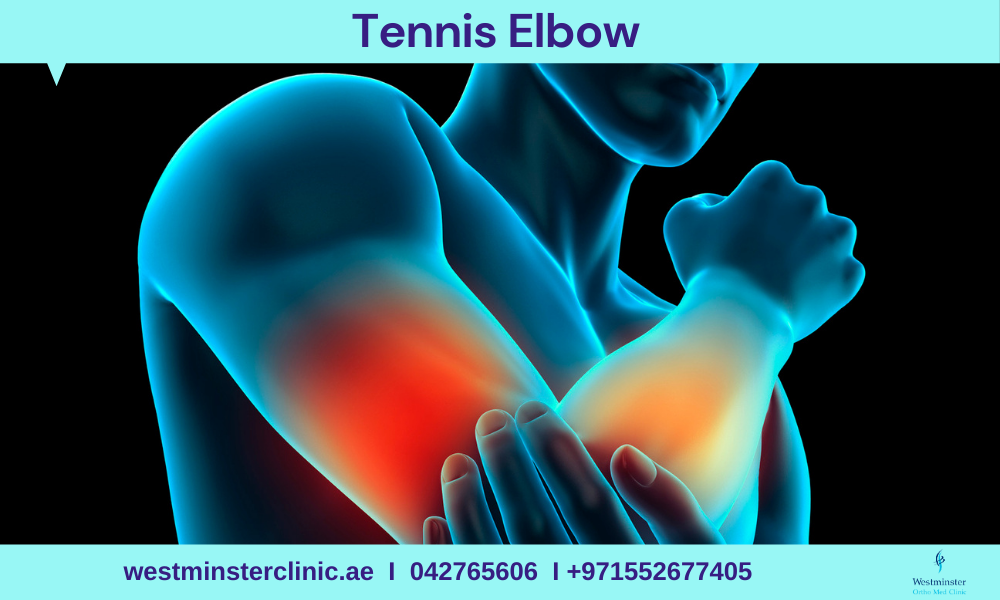
Tennis elbow, or lateral epicondylitis, is a painful condition of the elbow caused by overuse. Not surprisingly, playing tennis or other racquet sports can cause this condition. However, several other sports and activities besides sports can also put you at risk.

Tennis elbow is inflammation or, in some cases, microtearing of the tendons that join the forearm muscles on the outside of the elbow. The forearm muscles and tendons become damaged from overuse — repeating the same motions again and again. This leads to pain and tenderness on the outside of the elbow.

Causes of Tennis Elbow

Tennis elbow usually develops over time. Repetitive motions — like gripping a racket during a swing — can strain the muscles and put too much stress on the tendons. That constant tugging can eventually cause microscopic tears in the tissue.

Tennis elbow might result from:

  • Tennis
  • Racquetball
  • Squash
  • Fencing
  •  Weight lifting

It can also affect people with jobs or hobbies that require repetitive arm movements or gripping such as:

  • Carpentry
  • Typing
  • Painting
  • Raking
  • Knitting

Risk factors

Factors that may increase your risk of tennis elbow include:

  • Age. While tennis elbow affects people of all ages, it’s most common in adults between the ages of 30 and 50.
  • Occupation. People who have jobs that involve repetitive motions of the wrist and arm are more likely to develop tennis elbow. Examples include plumbers, painters, carpenters, butchers and cooks.
  • Certain sports. Participating in racket sports increases your risk of tennis elbow, especially if you employ poor stroke technique.

Symptoms of Tennis Elbow

The symptoms of tennis elbow include pain and tenderness in the bony knob on the outside of your elbow. This knob is where the injured tendons connect to the bone. The pain may also radiate into the upper or lower arm. Although the damage is in the elbow, you’re likely to hurt when doing things with your hands.

Tennis elbow is similar to another condition called golfer’s elbow, which affects the tendons on the inside of the elbow.

Tennis Elbow Treatment

Your doctor will first tell you to stop playing sports or doing certain kinds of work so your arm can rest. They’ll probably recommend physical therapy and some medications .

Your doctor will give you:

  • Corticosteroid medications. They’ll inject these into the area to help with swelling and pain.
  • A splint or brace. You’ll wear this on your arm to help your muscles and tendons rest.
  • Medications. Acetaminophen or anti-inflammatory medications may be taken to help reduce pain and swelling
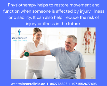
  • Physical therapy. Specific exercises are helpful for strengthening the muscles of the forearm. Your therapist may also perform ultrasound, ice massage, or muscle-stimulating techniques to improve muscle healing.
  • Ultrasound. This treatment can break up scar tissue, increase blood flow, and promote healing.
  • Brace. Using a brace centered over the back of your forearm may also help relieve symptoms of tennis elbow. This can reduce symptoms by resting the muscles and tendons.
  • Steroid injections. Steroids, such as cortisone, are very effective anti-inflammatory medicines. Your doctor may decide to inject the painful area around your lateral epicondyle with a steroid to relieve your symptoms.
  • Platelet-rich plasma. Platelet-rich plasma (PRP) is a biological treatment designed to improve the biologic environment of the tissue. This involves obtaining a small sample of blood from the arm and centrifuging it (spinning it) to obtain platelets from the solution.  Platelets are known for their high concentration of growth factors, which can be injected into the affected area. While some studies about the effectiveness of PRP have been inconclusive, others have shown promising results.

Up to 95% of people who have tennis elbow get better without surgery. But you might need it if you still have pain after 6 to 12 months. Your doctor can remove damaged tissue through cuts in your arm.

Tennis Elbow Prevention

Some small changes might help lower your risk of tennis elbow.

  • Keep your arms and wrists strong and flexible. Build strength with light weights. Warm up and stretch before any activity, especially one that involves making the same motions over and over.
  • Try not to make repetitive movements.
  • If you play a sport with a racquet, have an expert check your equipment to be sure it’s the right size and fit.

To reduce the risk of tennis elbow, it is important to pay attention to movement techniques during exercise or exertion.

It is better to spread the load to the larger muscles of the shoulder and upper arm, rather than focus activity on the smaller muscles in the wrist and the elbow.

Warming up: Warming up before playing a sport that involves repetitive arm movements, such as tennis or squash, is essential. Gently stretching the arm muscles will help to avoid injury.

Using lightweight tools: Lighter sports equipment or racquets with a larger grip size will help reduce strain on the tendons. Damp tennis balls and older balls load the arm with unnecessary force.

Increasing the strength of forearm muscles: This can help support arm movement and prevent tennis elbow.

A physical therapist can also recommend suitable exercises to help strengthen the appropriate muscles.

Should you require additional information or would like to make an appointment with our Consultant Orthopedic Surgeon Dr. Ishrat Khan OR Physical Therapists,  Anil Daniel,  OR Hadel Radwan  please call us or e-mail us at info@westminsterclinic.ae

Reference:

  • oxfordhealth.nhs.uk
  • yourphysio.org.uk
  • Mayoclinic.org

Disclaimer: All contents on this site are for general information and in no circumstances information be substituted for professional advice from the relevant healthcare professional, Writer does not take responsibility of any damage done by the misuse or use of the information.

January 13, 2021

An ankle sprain is an injury to the tough bands of tissue (ligaments) that surround and connect the bones around the ankle joint. It typically happens when you accidentally twist or turn your ankle in an awkward way. This can stretch or tear the ligaments but can also affect tendons and blood vessels in the surrounding areas.

Sprained Ankle Causes

Anything that stretches your ankle more than it’s used to can hurt a ligament. This usually happens when your foot is turned inward or twisted, such as when you:

  • Plant your foot the wrong way when running, stepping up or down, or doing everyday things like getting out of bed
  • Step on an uneven surface, like in a hole
  • Step on someone else while playing sports. (For example, your foot might roll when you’re playing basketball, go up for a rebound, and come down on top of another player’s foot.)

Sprained Ankle Symptoms

The inflammation that comes along with a sprained ankle can cause symptoms including:

  • Swelling
  • Tenderness
  • Bruising
  • Pain
  • Inability to put weight on the affected ankle
  • Skin discoloration
  • Stiffness

The ankle can sustain many different types of injuries. It’s important to see your doctor when you’re experiencing problems with your ankle. Your doctor can determine whether the injury is a sprain or something more severe.

When to call your doctor

You probably won’t need to see your doctor about a sprain. But give them a call if:

  • Your pain is severe or doesn’t get better with over-the-counter medications, elevation, and ice
  • You can’t walk, or you have severe pain when you do
  • Your ankle doesn’t feel better within 5 to 7 days

You might have a bone fracture instead of a sprain if:

  • You have severe pain or pain that doesn’t get better with treatment
  • Your foot or ankle is twisted or extremely swollen
  • You can’t walk without pain
  • You have severe pain when you press your medial malleolus or lateral malleolus, the bony bumps on each side of your ankle

Sprained Ankle Complications

Other problems can develop over time if you don’t treat a sprained ankle, try to do too much before it’s completely healed, or sprain it more than once. These complications include:

  • An unstable joint
  • Pain
  • Arthritis
  • Injuring the other ankle because of changes in how you walk

Ankle Sprain Treatment

Doctors recommend RICE:

  • Rest keeps you from hurting the ankle again or putting stress on inflamed tissue. A brace or splint can take pressure off the joint.
  • Ice is probably the best treatment. Put it on your ankle to lower blood flow and help with swelling, redness, and warmth. It can prevent inflammation if you do it quickly after an injury.
  • Compression can keep down swelling. Use an elastic bandage or wrap until the swelling goes down. Always start wrapping at the point farthest from your heart. Don’t wrap so tightly that you cut off the blood flow.
  • Elevation (keeping the injured area up as high as possible) will help your body absorb extra fluid. It’s best to prop your ankle up so that it’s higher than your heart, as with a reclining chair.

Anti-inflammatory pain medications reduce pain and fight swelling. Over-the-counter drugs work for most people. Check with your doctor first if you have other health conditions or take any other medicines.

How to do rehabilitation exercises for an ankle sprain

When the initial symptoms of pain and swelling start to subside, you will need to start some progressive exercise to ensure an optimal outcome. Below is an outline that may allow most people to return to their normal activities without any problems. Some who participate in sport involving changes of direction, jumping and landing may require more time or enhanced rehabilitation to achieve these goals.

  • Weight bearing and walking as early as possible
  • Maintain range of motion with some easy mobility exercises
  • Stretching exercises as Achilles tendon, Towel stretch and Calf stretch.
  • Basic strength exercise
  • Balance and control exercises
  • Gradually increase the distance you are walking and expose yourself to different types of terrain (incline/decline/uneven ground
  • Practice jumping and landing
  • Begin running and include changes of direction depending upon your rehabilitation goals.

In most cases you are able to walk relatively comfortably after 1-2 weeks, can use it fully after 6-8 weeks and return to sport after 12 weeks. This is however dependent upon how severe the sprain is.

Start each exercise slowly and use your pain level to guide you in doing these exercises. Ease off the exercise if you have more than mild pain. Following are some examples of typical rehabilitation (rehab) exercises.

Keep in mind that the timing and type of rehab exercises recommended for you may vary according to your doctor’s or physical therapist’s preferences.

How can I prevent an ankle sprain?

You can lower your risk for future sprains by:

  • wrapping the affected ankle in an elastic bandage
  • wearing a brace, if necessary
  • performing strengthening exercises
  • avoiding high heels
  • warming up before exercising
  • wearing sturdy, quality footwear
  • paying attention to surfaces you’re walking on
  • slowing or stopping activities when you feel fatigued

Call us right away if you think you’ve sprained your ankle again. When left untreated, an ankle sprain can lead to long-term pain and instability in the ankle.

Recovery


As your symptoms improve, gradually build up your walking times and ease back into normal activity.
1. Gentle exercise and getting back to normal will help the ligament heal quicker and stronger, continuing to rest it after the first few days will slow down the healing process.
2. It is not unusual for it to be a bit sore when you restart a new activity for the first time, so get back to your usual activities gently. It might help to ice it again after you do something new, for example, going to the gym for the
first time.
3. It usually takes six to eight weeks to get back to normal activity.
4. Physiotherapy can be very helpful in helping you maximize your recovery and reduce your risk of re-injury

Should you require additional information or would like to make an appointment with our Consultant Orthopedic Surgeon Dr. Ishrat Khan OR Physical Therapists,  Anil Daniel,  OR Hadel Radwan  please call us or e-mail us at info@westminsterclinic.ae

Disclaimer: All contents on this site are for general information and in no circumstances information be substituted for professional advice from the relevant healthcare professional, Writer does not take responsibility of any damage done by the misuse or use of the information.

Reference:

  • oxfordhealth.nhs.uk
  • yourphysio.org.uk
  • Harvard Medical School
  • Mayoclinic.org
January 12, 2021

Colorectal cancer starts in the colon or the rectum. These cancers can also be called colon cancer or rectal cancer, depending on where they start. Colon cancer and rectal cancer are often grouped together because they have many features in common. Cancer starts when cells in the body start to grow out of control.

The colon and rectum

To understand colorectal cancer, it helps to know about the normal structure and function of the colon and rectum.

The colon and rectum make up the large intestine (or large bowel), which is part of the digestive system, also called the gastrointestinal (GI) system (see illustration below).

Most of the large intestine is made up of the colon, a muscular tube about 5 feet (1.5 meters) long. The parts of the colon are named by which way the food is traveling through them.  

  • The first section is called the ascending colon. It starts with a pouch called the cecum, where undigested food is comes in from the small intestine. It continues upward on the right side of the abdomen (belly).
  • The second section is called the transverse colon. It goes across the body from the right to the left side.
  • The third section is called the descending colon because it descends (travels down) on the left side.
  • The fourth section is called the sigmoid colon because of its “S” shape. The sigmoid colon joins the rectum, which then connects to the anus. 

The ascending and transverse sections together are called the proximal colon. The descending and sigmoid colon are called the distal colon.

How do the colon and rectum work?

The colon absorbs water and salt from the remaining food matter after it goes through the small intestine (small bowel). The waste matter that’s left after going through the colon goes into the rectum, the final 6 inches (15cm) of the digestive system. It’s stored there until it passes through the anus. Ring-shaped muscles (also called a sphincter) around the anus keep stool from coming out until they relax during a bowel movement.

How does colorectal cancer start?

Polyps in the colon or rectum

Most colorectal cancers start as a growth on the inner lining of the colon or rectum. These growths are called polyps.

Some types of polyps can change into cancer over time (usually many years), but not all polyps become cancer. The chance of a polyp turning into cancer depends on the type of polyp it is. There are different types of polyps.

  • Adenomatous polyps (adenomas): These polyps sometimes change into cancer. Because of this, adenomas are called a pre-cancerous condition. The 3 types of adenomas are tubular, villous, and tubulovillous. 
  • Hyperplastic polyps and inflammatory polyps: These polyps are more common, but in general they are not pre-cancerous. Some people with large (more than 1cm) hyperplastic polyps might need colorectal cancer screening with colonoscopy more often. 
  • Sessile serrated polyps (SSP) and traditional serrated adenomas (TSA): These polyps are often treated like adenomas because they have a higher risk of colorectal cancer.

Other factors that can make a polyp more likely to contain cancer or increase someone’s risk of developing colorectal cancer include:

  • If a polyp larger than 1 cm is found
  • If more than 3 polyps are found
  • If dysplasia is seen in the polyp after it’s removed. Dysplasia is another pre-cancerous condition. It means there’s an area in a polyp or in the lining of the colon or rectum where the cells look abnormal, but they haven’t become cancer.

How colorectal cancer spreads

If cancer forms in a polyp, it can grow into the wall of the colon or rectum over time. The wall of the colon and rectum is made up of many layers. Colorectal cancer starts in the innermost layer (the mucosa) and can grow outward through some or all of the other layers (see picture below).

When cancer cells are in the wall, they can then grow into blood vessels or lymph vessels (tiny channels that carry away waste and fluid). From there, they can travel to nearby lymph nodes or to distant parts of the body.

Symptoms

The commonest symptom of colorectal cancer is a change of bowel habits. There may be increasing constipation, or perhaps alternating bouts of constipation and diarrhoea. There may be blood or mucus in the stools. A feeling that you haven’t completely emptied your bowels is quite common if the tumour is in the rectum. This can be uncomfortable and you may constantly feel the urge to go to the toilet.

You may feel a colicky type pain, or vague discomfort in your abdomen. You may also feel generally unwell, for example listless or tired, because you have been losing blood from the bowel and may have become anaemic (lack of red blood cells).

If your symptoms have lasted six or more weeks, including bleeding from the rectum, you need to see a specialist.

Colorectal cancer diagnosis

There are a series of tests and investigations which can be done to confirm or rule out a diagnosis of colorectal cancer, and to find out whether the cancer has spread to other parts of your body.

There are several ways which the doctor can examine your bowel. Whatever examination is used your bowel will need to be prepared. It must be as empty as possible so that the doctor can get a clear view inside.

The preparation may vary but will usually include:

  • Eating a low fibre (roughage) diet for a day or so before the test to clear your bowel of any residue
  • Drinking clear fluids only the day before the examination
  • Taking laxatives to clear your upper bowel
  • An enema to make sure the lower part of the bowel is empty.
  • If you have any questions or the explanation is unclear, please ask your doctor or nurse.

Sigmoidoscopy or colonoscopy

During these investigations the doctor passes a scope (a tube with a small camera on the end) into your bowel. A sigmoidoscopy looks at the lower part of your large bowel, while the colonoscopy looks further up the colon.

The doctor can see if there is part of the lining of the bowel which looks different, for example there may be a polyp (a small smooth growth) or an ulcer.

If the doctor does see something unusual, a biopsy (a sample of tissue) will be taken from this area. The tissue will be sent to the laboratory for examination under the microscope.

Before these examinations you will be given something to make you more relaxed and prevent any discomfort. If you have any questions, please ask your doctor or nurse.

Barium enema

This is an X-ray examination using barium which brightens the X-ray picture. The barium is given as an enema and will outline the lower part of your bowel.

The procedure lasts 15–30 minutes and you should try to hold the contents of the enema for the length of the examination. Afterwards you will be able to empty your bowels. You may be prescribed a mild laxative because barium can cause constipation. Barium can also be very difficult to flush away in the toilet. If you have any questions, please ask your doctor or nurse.

Other tests

You may have other tests, which can include blood tests, abdominal ultrasound, CT scan or MRI scan.

The information from these tests is used to assess the size of the cancer and how far it has spread. This is called ‘staging’. Your doctors need to know the extent of the cancer to help them decide on the most appropriate treatment for you.

Staging of colorectal cancer is based on a physical examination, the results of tests and what the doctors find at the time of surgery.

Colorectal cancer treatment

Treatment will usually be an operation to remove the cancer and/or to relieve your symptoms. You may also have chemotherapy or radiotherapy as well as an operation.

There are several ways of treating colorectal cancer and your treatment will be planned individually for you. Don’t be concerned if you talk to other people who are receiving similar, but different, treatments.

Surgery

An operation may be performed to remove the cancer and part of the bowel on either side of this. Depending on the extent of the operation the two ends of the bowel may be stitched together.

If the tumour is sited low in rectum, there may not be enough bowel left to join together. In this case you may have to have a colostomy formed. A colostomy, or stoma, is an artificial opening created when the healthy part of your bowel is brought out onto the surface of your abdomen. Your stool will be passed through this opening instead of through your rectum as before. You will need to wear an appliance (bag) to collect your stools. This type of colostomy will be permanent.

In some situations you may need to have a temporary colostomy formed to rest of the bowel while healing takes place. This is usually only for a few weeks and will be discussed with you by your doctor.

If it is possible to say before your operation that you will need to have a colostomy, a stoma care nurse will visit you. They will explain exactly what will happen and what to expect. The stoma care nurse will show you how to care for your colostomy and help you adapt to living with a stoma. They can provide support over a long period of time.

If you have any questions or there is anything you don’t understand, please speak to your doctor or nurse.

Chemotherapy

Chemotherapy (drug treatment) may be recommended for you. Chemotherapy means treatment with anti-cancer drugs, which are given to destroy or control cancer cells by damaging them so that they can’t divide and grow.

Radiotherapy

Radiotherapy uses high-energy X-rays to kill cancer cells and is given using a machine similar to an X-ray machine but slightly larger. The treatment area will include the tumour and the surrounding lymph nodes (glands) if necessary. The treatment will planned specifically for you to make sure that the cancer cells are destroyed with the least amount of damage to normal tissues.

Side effects

Your bowel habits may change during radiotherapy, for example you stool may become loose or you may develop diarrhoea. If this happens, please tell your doctor, radiographer or nurse. You will be given advice on diet and medicine can be prescribed to help you. Make sure you drink plenty of fluids.

Your bladder may be included in the treatment field and, if so, you may experience some discomfort when passing urine. You may also want to pass urine more frequently. Once again, make sure you drink plenty of fluids and tell your doctor about this problem.

After treatment for colorectal cancer

Having and being treated for colorectal cancer will have an effect on your life, and there are other things which you should be aware of in addition to the side effects of treatment.

Checkups

The doctor will ask you to attend hospital at regular intervals during and after your treatment. You will be given an appointment for the outpatients clinic and, as time passes, the appointments will probably become less frequent.

Each time you attend, the doctors will examine you. Blood tests or X-rays may be repeated to check your recovery and make sure the cancer hasn’t come back.

Eating and drinking: After treatment for colorectal cancer there are usually no restrictions on what you can eat and drink, including alcohol in moderation. If you would like advice about your diet, please ask to see the dietician.

Should you require additional information OR would like to make an appointment with our Consultant Colorectal Surgeon, Dr. Majid Bassuni , please call us or e-mail us at info@westminsterclinic.ae

Disclaimer: All contents on this site are for general information and in no circumstances information be substituted for professional advice from the relevant healthcare professional, Writer does not take responsibility of any damage done by the misuse or use of the information.

Reference:

  • NHS.uk
  • American Cancer Society
  • Harvard Medical School
  • Mayoclinic.org

January 11, 2021

Osteoarthritis of the knee occurs when the bony surfaces (cartilage) between the thigh bone and shin bone, and sometimes on the back of the kneecap (patella), become worn and uneven. This prevents smooth movement of the joint and can be a normal part of the ageing process.

Knee osteoarthritis (knee OA) is a progressive disease caused by inflammation and degeneration of the knee joint that worsens over time. It affects the entire joint, including bone, cartilage, ligaments, and muscles. Its progression is influenced by age, body mass index (BMI), bone structure, genetics, muscular strength, and activity level. Knee OA also may develop as a secondary condition following a traumatic knee injury. Depending on the stage of the disease and whether there are associated injuries or conditions, knee OA can be managed with physical therapy. More severe or advanced cases may require surgery.


Symptoms

  • Pain and swelling
  • Stiffness and restricted movement
  • Grinding noise on movement
  • The knee giving way when turning or walking on uneven ground
  • Pain first thing in the morning, after Sitting too long or after doing too much.
  • Tenderness –  Your joint might feel tender when you apply light pressure to or near it.
  • Loss of flexibility –  You might not be able to move your joint through its full range of motion.
  • Bone spurs –  These extra bits of bone, which feel like hard lumps, can form around the affected joint.

Managing osteoarthritis of the knee

1. Weight loss: Lightening the load

Every step you take exposes your knee joints to a force equal to three to five times your body weight. If you have osteoarthritis, a weight gain or loss of just 5 pounds can cause a noticeable difference in the amount of pain you feel. One study of overweight or obese older adults who had knee osteoarthritis found that for every pound of weight they lost, the stress on their knees was reduced fourfold. Obesity not only puts added stress on the knees, it also spurs the production of inflammatory proteins that may hasten cartilage degeneration. Weight loss can help preserve cartilage and reduce symptoms.

2. Exercise: Motion as medicine

Lack of exercise and knee osteoarthritis can produce a downward spiral of increasing disability. Lack of exercise makes you more likely to develop knee osteoarthritis; the pain of osteoarthritis makes you avoid exercise; and avoiding exercise makes the arthritis worse. The key to breaking this cycle is exercise.

Regular light to moderate exercise (during which you sweat lightly but can talk easily) can slow the disease process and reduce your pain. The exercise should be individually tailored to prevent injury, so start with an evaluation by a clinician or physical therapist experienced in managing osteoarthritis. A program to improve knee osteoarthritis may include the following:

Low-impact aerobic exercise . Swim, cycle, walk, or use an elliptical trainer (a machine that simulates walking or stair-climbing without stressing the joints) and gradually increase the time you spend doing it. Also, try to incorporate more activity in your daily tasks—for example, park farther from your destination and walk, or use stairs instead of the elevator.

Quadriceps strengthening . Strengthening the thigh muscles will help protect the knee and improve pain, stiffness, and balance. (For exercises that work the quadriceps, see “Knee-strengthening exercises”) If your joints are poorly aligned or the ligaments are overstretched, consult a physical therapist for safe strengthening exercises.

Flexibility . Muscle stiffness can limit knee-joint movement and lead to further pain. Stretching and range-of-motion exercises may help.

Balance . Knee osteoarthritis can interfere with balance by impairing the capacity of receptors in your joints to detect the position of your body in space. You can improve your balance with strengthening exercises as well as specific balance exercises and activities such as tai chi.

3. Reduce pain with medications

Medications can’t change the course of osteoarthritis, but they can help ease pain and make it possible for you to exercise.

How Can a Physical Therapist Help?

Once you have received a diagnosis of Osteoarthritis of the knee , your physical therapist will design an individualized treatment program specific to the exact nature of your condition and your goals. Your treatment program may include:

  • Range-of-motion exercises.
  • Muscle strengthening. 
  • Manual therapy.
  • Bracing.
  • Activity recommendations.
  • Modalities.

Regular contact with a health professional will help you make the most of self-management strategies, medications, and other means of slowing the course of knee osteoarthritis and maintaining the best possible quality of life.

Should you require additional information or would like to make an appointment with our Consultant Orthopedic Surgeon Dr. Ishrat Khan OR Physical Therapists,  Anil Daniel,  OR Hadel Radwan  please call us or e-mail us at info@westminsterclinic.ae

Disclaimer: All contents on this site are for general information and in no circumstances information be substituted for professional advice from the relevant healthcare professional, Writer does not take responsibility of any damage done by the misuse or use of the information.

Reference:

  • NHS.uk
  • Physio.co.uk
  • Harvard Medical School
  • Mayoclinic.org

January 10, 2021

Obsessive-compulsive disorder (OCD) has two main parts: obsessions and compulsions.

  • Obsessions are unwelcome thoughts, images, urges, worries or doubts that repeatedly appear in your mind. They can make you feel very anxious (although some people describe it as ‘mental discomfort’ rather than anxiety).
  • Compulsions are repetitive activities that you do to reduce the anxiety caused by the obsession. It could be something like repeatedly checking a door is locked, repeating a specific phrase in your head or checking how your body feels.

It’s not about being tidy, it’s about having no control over negative thoughts. It’s about being afraid not doing things a certain way will cause harm.”

Person with OCD might find that sometimes the obsessions and compulsions are manageable, and at other times they may make day-to-day life really difficult. They may be more severe when one stressed about other things, like life changes, health, money, work or relationships.

What’s it like to live with OCD?

Although many people experience minor obsessions (such as worrying about leaving the gas on, or if the door is locked) and compulsions (such as avoiding the cracks in the pavement), these don’t significantly interfere with daily life, or are short-lived.

If you experience OCD, it’s likely that your obsessions and compulsions will have a big impact on how you live your life:

  • Disruption to your day-to-day life. Repeating compulsions can take up a lot of time, and you might avoid certain situations that trigger your OCD. This can mean that you’re not able to go to work, see family and friends, eat out or even go outside. Obsessive thoughts can make it hard to concentrate and leave you feeling exhausted.
  • Impact on your relationships. You may feel that you have to hide your OCD from people close to you – or your doubts and anxieties about a relationship may make it too difficult to continue.
  • Feeling ashamed or lonely. You may feel ashamed of your obsessive thoughts, or worry that they can’t be treated. You might want to hide this part of you from other people, and find it hard to be around people or to go outside. This can make you feel isolated and lonely.
  • Feeling anxious. You may find that your obsessions and compulsions are making you feel anxious and stressed. For example, some people feel that they become slaves to their compulsions and have to carry them out so frequently that they have little control over them.

What causes OCD?

There are different theories about why OCD develops. None of these theories can fully explain every person’s experience, but researchers suggest that these are likely to be involved in causing OCD:

  1. Personal experience

Some theories suggest that OCD is caused by personal experience. For example:

  • If you’ve had a painful childhood experience, or suffered trauma, abuse or bullying, you might learn to use obsessions and compulsions to cope with anxiety.
  • If your parents had similar anxieties and showed similar kinds of compulsive behavior, you may have learned OCD behaviors as a coping technique.
  • Ongoing anxiety or stress, or being part of a stressful event like a car accident or starting a new job, could trigger OCD or make it worse.
  • Pregnancy or giving birth can sometimes trigger perinatal OCD. 

2. Personality

Some research suggests that people with certain personality traits may be more likely to have OCD. For example, if you are a neat, meticulous, methodical person with high standards, you may be more likely to develop OCD.

3. Biological factors

Some biological theories suggest that a lack of the brain chemical serotonin may have a role in OCD. However, it’s unclear this is the cause or is an effect of the condition.

Studies have also looked at genetic factors and how different parts of the brain might be involved in causing OCD, but have found nothing conclusive.

Symptoms of obsessive compulsive disorder (OCD)

If you have OCD, you’ll usually experience frequent obsessive thoughts and compulsive behaviours.

  • An obsession is an unwanted and unpleasant thought, image or urge that repeatedly enters your mind, causing feelings of anxiety, disgust or unease.
  • A compulsion is a repetitive behaviour or mental act that you feel you need to do to temporarily relieve the unpleasant feelings brought on by the obsessive thought.

For example, someone with an obsessive fear of being burgled may feel they need to check all the windows and doors are locked several times before they can leave their house. Women can sometimes have OCD during pregnancy or after their baby is born. Obsessions may include worrying about harming the baby or not sterilizing feeding bottles properly. Compulsions could be things such as repeatedly checking the baby is breathing.

Treatments for obsessive compulsive disorder (OCD)

There are some effective treatments for OCD that can help reduce the impact it has on your life.

The main treatments are:

  • Psychological therapy – usually cognitive behavioural therapy (CBT) which helps you face your fears and obsessive thoughts without “putting them right” through compulsions
  • Medicine – usually a type of antidepressant medicine called selective serotonin reuptake inhibitors (SSRIs) which can help by altering the balance of chemicals in your brain.

CBT will usually have an effect quite quickly. It can take several months before you notice the effects of treatment with SSRIs, but most people will eventually benefit.

If these treatments do not help, you may be offered an alternative SSRI or be given a combination of an SSRI and CBT. Some people may be referred to a specialist mental health service for further treatment.

Getting help for Obsessive Compulsive Disorder (OCD)

Obsessions and compulsions can take over your life, and leave you feeling helpless. However, there are some things you can try to help manage your OCD and improve your wellbeing.

Remember that different things work for different people at different times. If something isn’t working for you (or doesn’t feel possible just now), you can try something else or come back to it another time.

  1. Self-help resources

Self-help resources for OCD are designed to help you develop coping strategies and are often based on cognitive behavioural therapy (CBT). Refer yourself directly to a psychological therapies service – find a psychological therapies service in your area

2. Build Your Support Network

Many people find it hard to talk about OCD. You might worry that people won’t understand. You might have kept your OCD secret for such a long time that it feels very scary to put some of your experiences into words. Strengthening the relationships around you may help you feel less lonely and more able to cope.

  • Talk to someone you trust about your OCD. Find a quiet space to talk where you won’t be interrupted. Some people find it helpful to write their feelings down and then talk about this together.
  • Spend time with friends and family. You might not feel ready to talk openly about your OCD yet. But spending more time with friends and family may help you feel more comfortable around them and, in time, more able to share your experiences.

3. Try Peer Support

Making connections with people with similar or shared experiences can be really helpful. You could try talking to other people who have OCD to share your feelings, experiences and ideas for looking after yourself. For example, you could:

  • Contact your doctor for group support
  • Try an online peer support community.
  • Find a local support group through an organisation such as OCD

4. Learn to Let Go

  • Manage your stress. Stress and anxiety can make OCD worse. You can read more about how to manage stress here.
  • Try a relaxation technique. Relaxation can help you look after your wellbeing when you are feeling stressed, anxious or busy. You can read more about relaxation here.
  • Try mindfulness. You might find that your CBT therapist includes some principles of mindfulness in your therapy. Mindfulness can help you reduce stress and anxiety. For some people, it can be helpful as part of recovery from OCD.

5. Look after your physical health

  • Get enough sleep. Sleep can give you the energy to cope with difficult feelings and experiences.
  • Think about your diet. Eating regularly and keeping your blood sugar stable can make a difference to your mood and energy levels. You can read more about food and mental health here.
  • Try to do some physical activity. Many people find exercise a challenge but activities like yoga, swimming or walking can help improve your mood. Any kind of physical activity counts – from a chair-based exercise regime to dancing round the kitchen – the important thing is to find something that works for you. You can read more about physical activity and mental health here.

Should you require additional information or would like to make an appointment with our Psychiatrist  please call us or e-mail us at info@westminsterclinic.ae

Reference:

  • NHS.uk
  • mind.org.uk
  • Johns Hopkins Medicine

Disclaimer: All contents on this site are for general information and in no circumstances information be substituted for professional advice from the relevant healthcare professional, Writer does not take responsibility of any damage done by the misuse or use of the information.

January 9, 2021

Shoulder impingement syndrome is the result of a vicious cycle of rubbing of the rotator cuff between your humerus and top outer edge of your shoulder. The rubbing leads to more swelling and further narrowing of the space, which result in pain and irritation. Treatments include rest, ice, anti-inflammatory medications, physical therapy, cortisone injections and surgery.

How does shoulder impingement syndrome develop?

When your rotator cuff is irritated or injured, it swells in much the same way your ankle does when it is sprained. However, because your rotator cuff is surrounded by bone, swelling causes other events to occur. Swelling reduces the amount of space around the rotator cuff, leading to rubbing against the acromion. Like a vicious cycle, the rubbing of the rotator cuff tendons result in swelling, which further narrows the space below the acromion. In some cases, bone spurs on the acromion bone can contribute to impingement by causing the space where the rotator cuff sits to be even more narrowed

What are the symptoms of shoulder impingement?

The first symptoms of impingement are mild. Therefore, many individuals do not seek treatment straight away. Symptoms gradually worsen over time. Initial symptoms include:

  • Reduced range of movement
  • Swelling/ inflammation
  • Stiffness
  • Weakness
  • Joint clicking
  • Joint locking

In advanced cases, impingement may progress to a “frozen Shoulders .

How is shoulder impingement syndrome diagnosed?

Your healthcare provider will take your medical history and perform a physical examination to check for pain and tenderness. Your provider will also assess the range of motion of your shoulder and your arm/shoulder strength. They will ask if you are involved in repetitive activities that require overhead arm movement, what remedies you have tried to relieve your pain, prior injuries to the affected arm/shoulder and any details about what may have caused your shoulder pain and when it began.

X-rays are helpful to rule out arthritis and may show the rotator cuff injury. There may be bone spurs or changes in the normal outline of the bone where the rotator cuff normally attaches. Magnetic resonance imaging (MRI ) and ultrasound can show tears in the rotator cuff tendons and inflammation in the bursa.

A diagnosis of impingement syndrome may be made if a small amount of an anesthetic (painkiller), injected into the space under the acromion, relieves your pain.

Management and Treatment

The goal of treatment for shoulder impingement syndrome is to reduce your pain and restore shoulder function. Treatments for impingement syndrome include rest, ice, over-the-counter anti-inflammatory medications, injections and physical therapy.

  • Physical therapy is the most important treatment for shoulder impingement syndrome. In most cases, you’ll only need one or two in-office visits to learn how to continue to do physical therapy on your own at home. You’ll learn stretching exercises to improve the range of motion of your shoulder. As your pain lessens, you can begin strengthening exercises to improve your rotator cuff muscles.
  • Ice should be applied to the shoulder for 20 minutes once or twice a day. (A bag of frozen peas or corn also works well).
  • If your healthcare provider approves, ibuprofen or naproxen may be taken as-needed to relieve pain. For more severe pain, a stronger prescription strength anti-inflammatory medication may be prescribe or a cortisone injection into the bursa beneath the acromion may be given.
  • A common sense approach to activities is helpful. Avoid activities in which you need to frequently reach overhead or behind your back. These motions usually makes shoulders with impingement syndrome worse. Stop activities that involve these motions until your pain improves.
  • Surgery is considered if nonsurgical treatments don’t relieve your pain. One surgery called a subacromial decompression or arthroscopic shoulder decompression removes part of the acromion to create more space for the rotator cuff. This procedure can be performed arthroscopically, which means through small cuts (incisions) in your shoulder. Other problems with your shoulder can be repaired at the time of surgery including arthritis in the shoulder, inflammation of the biceps tendon and partial rotator cuff tear. Open surgery, a surgery with a larger cut in the front of your shoulder, is another option.

Should you require additional information or would like to make an appointment with our Consultant Orthopedic Surgeon Dr. Ishrat Khan OR Physical Therapists,  Anil Daniel,  OR Hadel Radwan  please call us or e-mail us at info@westminsterclinic.ae

Disclaimer: All contents on this site are for general information and in no circumstances information be substituted for professional advice from the relevant healthcare professional, Writer does not take responsibility of any damage done by the misuse or use of the information.

Reference:

  • NHS.uk
  • Physio.co.uk
  • Clevelandclinic.org
  • Johns Hopkins Medicine

January 7, 2021

Chronic pain is defined as pain that lasts at least 12 weeks. The pain may feel sharp or dull, causing a burning or aching sensation in the affected areas. It may be steady or intermittent, coming and going without any apparent reason. Chronic pain can occur in nearly any part of your body. The pain can feel different in the various affected areas.

Most common types of chronic pain include:

  • Headache
  • Postsurgical pain
  • Post-trauma pain
  • Lower back pain
  • Cancer pain
  • Arthritis pain
  • Neurogenic pain (pain caused by nerve damage)
  • Psychogenic pain (pain that isn’t caused by disease, injury, or nerve damage)

Causes of Chronic Pain?

Chronic pain is usually caused by an initial injury, such as a back sprain or pulled muscle. It’s believed that chronic pain develops after nerves become damaged. The nerve damage makes pain more intense and long lasting. In these cases, treating the underlying injury may not resolve the chronic pain.

In some cases, however, people experience chronic pain without any prior injury. The exact causes of chronic pain without injury aren’t well understood. The pain may sometimes result from an underlying health condition, such as:

  • Chronic fatigue syndrome: characterized by extreme, prolonged weariness that’s often accompanied by pain
  • Endometriosis: a painful disorder that occurs when the uterine lining grows outside of the uterus
  • Fibromyalgia: widespread pain in the bones and muscles
  • Inflammatory bowel disease: a group of conditions that causes painful, chronic inflammation in the digestive tract
  • Interstitial cystitis: a chronic disorder marked by bladder pressure and pain
  • Temporomandibular joint dysfunction (TMJ): a condition that causes painful clicking, popping, or locking of the jaw.

Chronic Pain Treatment

The main goal of treatment is to reduce pain and boost mobility. This helps you return to your daily activities without discomfort.

The severity and frequency of chronic pain can differ among individuals. So doctors create pain management plans that are specific to each person. Your pain management plan will depend on your symptoms and any underlying health conditions. Medical treatments, lifestyle remedies, or a combination of these methods may be used to treat your chronic pain.

Medications for chronic pain

Several types of medications are available that can help treat chronic pain. Here are a few examples:

  • Over-the-counter pain relievers.
  • Opioid pain relievers
  • Adjuvant analgesics

Medical procedures for chronic pain

Certain medical procedures can also provide relief from chronic pain. An example of a few are:

  • Electrical stimulation, which reduces pain by sending mild electric shocks into your muscles
  • Nerve block, which is an injection that prevents nerves from sending pain signals to your brain
  • Acupuncture, which involves lightly pricking your skin with needles to alleviate pain
  • Surgery, which corrects injuries that may have healed improperly and that may be contributing to the pain

Lifestyle remedies for chronic pain

Additionally, various lifestyle remedies are available to help ease chronic pain. Examples include:

  • Physical therapy
  • Tai chi
  • Yoga
  • Art and music therapy
  • Pet therapy
  • Psychotherapy
  • Massage
  • Meditation

Dealing with chronic pain

There isn’t a cure for chronic pain, but the condition can be managed successfully. It’s important to stick to your pain management plan to help relieve symptoms.

Physical pain is related to emotional pain, so chronic pain can increase your stress levels. Building emotional skills can help you cope with any stress related to your condition. Here are some steps you can take to reduce stress:

Take good care of your body: Eating well, getting enough sleep, and exercising regularly can keep your body healthy and reduce feelings of stress.

Continue taking part in your daily activities: You can boost your mood and decrease stress by participating in activities you enjoy and socializing with friends. Chronic pain may make it challenging to perform certain tasks. But isolating yourself can give you a more negative outlook on your condition and increase your sensitivity to pain.

Seek support: Friends, family, and support groups can lend you a helping hand and offer comfort during difficult times. Whether you’re having trouble with daily tasks or you’re simply in need of an emotional boost, a close friend or loved one can provide the support you need.

Should you require additional information or would like to make an appointment with our Consultant , Dr. Tarek Sultan,  please call us or e-mail us at info@westminsterclinic.ae

Disclaimer: All contents on this site are for general information and in no circumstances information be substituted for professional advice from the relevant healthcare professional, Writer does not take responsibility of any damage done by the misuse or use of the information.

Reference:

  • American Chronic Pain Association
  • American Pain Society
  • Health.harvard.edu

January 7, 2021

Cervical vertigo, or cervicogenic dizziness, is a neck-related sensation in which a person feels like either they’re spinning or the world around them is spinning. Poor neck posture, neck disorders, or trauma to the cervical spine cause this condition. 

Causes of cervical vertigo

There are a number of potential causes of cervical vertigo, though this condition is still being researched.

Blockage of arteries in the neck from hardening (atherosclerosis) or tearing of these arteries (dissection) are causes. The dizziness is caused in these cases by a disruption of blood flow to the inner ear or to a lower brain region called the brain stem. Arthritis, surgery, and trauma to the neck can also block blood flow to these important regions, resulting in this type of vertigo.

Cervical spondylosis (advanced neck osteoarthritis) may be another potential cause of neck-related dizziness. This condition causes your vertebrae and neck disks to wear and tear over time. This is called degeneration, and it can put pressure on the spinal cord or spinal nerves and block blood flow to the brain and inner ear. A slipped disk alone (herniated) can do the same thing without any spondylosis.

The muscles and joints in your neck have receptors that send signals about head movement and orientation to the brain and vestibular apparatus — or parts of the inner ear responsible for balance. This system also works with a larger network in the body to maintain balance and muscle coordination. When this system works improperly, receptors can’t communicate to the brain and cause dizziness and other sensory dysfunctions.

Symptoms of Cervical Vertigo

Cervical vertigo is associated with dizziness from sudden neck movement, specifically from turning your head. Other symptoms of this condition include:

  • Headache
  • Nausea
  • Vomiting
  • Ear pain or ringing
  • Neck pain
  • Loss of balance while walking, sitting, or standing
  • Weakness
  • Problems concentrating

Dizziness from cervical vertigo can last minutes or hours. If neck pain decreases, the dizziness may also begin to subside. Symptoms may worsen after exercise, rapid movement and sometimes sneezing.

Diagnosis

Diagnosing cervical vertigo can be difficult. Doctors will have to eliminate other potential causes of cervical vertigo with similar symptoms, including:

  • Benign positional vertigo
  • Central vertigo, which can be due to stroke, tumors, or multiple sclerosis
  • Psychogenic vertigo
  • Inner ear diseases, such as vestibular neuronitis

Once other causes and conditions are ruled out, doctors will perform a physical examination that requires turning your head. If there is sporadic eye movement (nystagmus) based on head positioning, you may have cervical vertigo.

Additional tests to confirm this diagnosis may include:

  • MRI scan of the neck
  • Magnetic resonance angiography (MRA)
  • Vertebral Doppler ultrasound
  • Vertebral angiography
  • Flexion-extension X-ray of the cervical spine
  • Evoked potential tests, which measure spinal cord and brain pathways in the nervous system

Cervical Vertigo Treatment

Treating cervical vertigo depends on treating the underlying cause. If you’re experiencing neck pain or have a degenerative neck disease, follow your medical treatment plan to decrease vertigo symptoms.

Doctors may also prescribe medication to reduce neck tightness, dizziness, and pain symptoms. Common medications prescribed include:

  • Muscle relaxants
  • Analgesics
  • Anti-dizziness drugs

Doctors also recommend physical therapy to improve your neck’s range of motion and your balance. Stretching techniques, therapy, and training on proper posture and use of your neck help to improve this condition. In some cases, where there is no risk to the patient, chiropractic manipulation of your neck and spine and heat compresses may decrease symptoms.

Outlook

Cervical vertigo is a treatable condition. Without proper medical guidance, your symptoms could get worse. Self-diagnosis is not recommended since this condition can mimic more serious diseases.

If you begin to experience dizziness, neck pain, and other related symptoms, visit your doctor immediately.

Disclaimer: All contents on this site are for general information and in no circumstances information be substituted for professional advice from the relevant healthcare professional, Writer does not take responsibility of any damage done by the misuse or use of the information.

January 3, 2021

Physiotherapy helps to restore movement and function when someone is affected by injury, illness or disability. It can also help to reduce the risk of injury or illness in the future.

When is physiotherapy used?

Physiotherapy can be helpful for people of all ages with a wide range of health conditions, including problems affecting the:

  • Bones, joints and soft tissue – such as back pain, neck pain, shoulder pain and sports injuries
  • Brain or nervous system – such as movement problems resulting from a stroke, multiple sclerosis (MS) or Parkinson’s disease
  • Heart and circulation – such as rehabilitation after a heart attack
  • Lungs and breathing – such as chronic obstructive pulmonary disease (COPD) and cystic fibrosis

Physiotherapy can improve your physical activity while helping you to prevent further injuries.

What physiotherapists do

Physiotherapists consider the body as a whole, rather than just focusing on the individual aspects of an injury or illness.

Some of the main approaches used by physiotherapists include:

  • Education and advice – physiotherapists can give general advice about things that can affect your daily lives, such as posture and correct lifting or carrying techniques to help prevent injuries
  • Movement, tailored exercise and physical activity advice – exercises may be recommended to improve your general health and mobility, and to strengthen specific parts of your body
  • Manual therapy – where the physiotherapist uses their hands to help relieve pain and stiffness, and to encourage better movement of the body.

Physiotherapy Techniques

Physiotherapy can involve a number of different treatment and preventative approaches, depending on the specific problems you’re experiencing.

At your first appointment, you will have an assessment to help determine what help you might need.

Three of the main approaches a physiotherapist may use are:

1. Education and advice:

One of the main aspects of physiotherapy involves looking at the body as a whole, rather than focusing on the individual factors of an injury. Therefore, giving general advice about ways to improve your wellbeing – for example, by taking regular exercise and maintaining a healthy weight for your height and build – is an important part of treatment.

A physiotherapist can also give you specific advice that you can apply to everyday activities to look after yourself and reduce your risk of pain or injury.

For example, if you have back pain, you may be given advice about good posture, correct lifting or carrying techniques, and avoiding awkward twisting, over-stretching or prolonged standing.

2. Movement and exercise:

Physiotherapists usually recommend movement and exercise to help improve your mobility and function. This may include:

  • Exercises designed to improve movement and strength in a specific part of the body – these usually need to be repeated regularly for a set length of time
  • Activities that involve moving your whole body, such as walking or swimming – these can help if you’re recovering from an operation or injury that affects your mobility
  • Exercises carried out in warm, shallow water (hydrotherapy or aquatic therapy) – the water can help relax and support the muscles and joints, while providing resistance to help you gradually get stronger
  • Advice and exercises to help you increase or maintain your physical activity – advice will be given on the importance of keeping active, and how to do this in a safe, effective way
  • Providing mobility aids – such as crutches or a walking stick to help you move around.

Your physiotherapist may also recommend exercises that you can continue doing to help you manage pain in the long term or reduce your risk of injuring yourself again.

3. Manual therapy:

Manual therapy is a technique where a physiotherapist uses their hands to manipulate, mobilise and massage the body tissues.

This can help:

  • Relieve pain and stiffness
  • Improve blood circulation
  • Help fluid drain more efficiently from parts of the body 
  • Improve the movement of different parts of the body 
  • Promote relaxation

Manual therapy can be used to treat specific problems, such as back pain, but may also be useful for a range of conditions that don’t affect the bones, joints or muscles.

For example, massage may improve quality of life for some people with serious or long-term conditions by reducing levels of anxiety and improving sleep quality. Manual techniques are also used to help certain lung conditions.

Other techniques:

Other techniques sometimes used by physiotherapists that may help to ease pain and promote healing include:

  • Acupuncture – where fine needles are inserted into specific points of the body, with the aim of reducing pain and promoting recovery
  • Transcutaneous electrical nerve stimulation (TENS) – a small, battery-operated device is used to deliver an electric current to the affected area, with the aim of relieving pain
  • Ultrasound – where high-frequency sound waves are used to treat deep tissue injuries by stimulating blood circulation and cell activity, with the aim of reducing pain and spasms, as well as speeding up healing

Some people have found these treatments effective, but there isn’t much scientific evidence to support them. There is some positive evidence for acupuncture, but the National Institute for Health and Care Excellence (NICE) only recommends considering it for chronic tension-type headaches and migraines.

Should you require additional information or would like to make an appointment with our Physical Therapists,  Anil Daniel,  OR Hadel Radwan  please call us or e-mail us at info@westminsterclinic.ae

Disclaimer: All contents on this site are for general information and in no circumstances information be substituted for professional advice from the relevant healthcare professional, Writer does not take responsibility of any damage done by the misuse or use of the information.

Reference:

  • NHS.uk
  • Johns Hopkins Medicine

January 2, 2021

The liver is the largest organ in the human body. It performs over 500 functions, including digestion of proteins, mineral storage, bile production and blood filtration. Many health problems can keep your liver from functioning properly and cause disease. There are several common symptoms of liver disease, including jaundice, liver failure and liver enlargement.

Liver Function

The liver performs many functions essential for good health and long life. Among its most important jobs are:

  • Producing important substances. Your liver continually produces bile. This is a chemical that helps turn fats into energy that your body uses. Bile is necessary for the digestive process. Your liver also creates albumin. This is a blood protein that helps carry hormones, drugs and fatty acids throughout your body. Your liver also creates most of the substances that help your blood clot after injury.
  • Processing bilirubin. The liver helps your body get rid of bilirubin, a substance found in bile. This happens from the breakdown of your red blood cells. Too much bilirubin in your body can cause jaundice. This is a yellowing of the skin and eyes.
  • Removing waste products. When you take in a potentially toxic substance, like alcohol or medicine, your liver helps alter it and remove it from your body.
  • Controlling immune responses. When bacteria, viruses and other harmful organisms enter your body, specialized cells in your liver can find and destroy these organisms.
  • Maintaining glucose. The liver helps your body maintain a healthy level of blood sugar. Your liver supplies glucose to your blood when it’s needed. It also removes glucose from your blood when there’s too much.

Types of liver disease:

  • Alcoholic Liver Disease. Alcoholic liver disease is a result of alcohol abuse. A large percentage of Americans drink alcohol, and most do not develop liver disease as a result. However, those who continue to consume alcohol excessively may cause injury to their liver.
  • Cholestasis. This happens when the flow of bile from your liver is limited or blocked. Cholestasis can be caused by certain drugs, genetic factors or even pregnancy. It can also happen from a blockage caused by a tumor or a gallstone stuck in the body’s digestive system.
  • Cirrhosis. This is a hardening of your liver due to scar tissue. Heavy alcohol use and viruses like hepatitis are common causes of cirrhosis. Diabetes, immune problems and genetic diseases can also cause the disease.
  • Hepatitis. This is the name for any condition involving inflammation of your liver. There are many different types. Sometimes, excessive alcohol use, drugs or toxins cause hepatitis. Hepatitis can lead to liver failure, liver cancer and other life-threatening conditions.
  • Nonalcoholic Fatty Liver Disease. This happens when there are fat deposits in the liver. The deposits prevent your liver from functioning properly and removing toxins from your body.

Symptoms of liver disease

Most types of liver disease do not cause any symptoms in the early stages. Once you start to get symptoms of liver disease, your liver is already damaged and scarred. This is known as cirrhosis.

See your Doctor if you have symptoms of liver damage (cirrhosis), such as:

  • Feeling very tired and weak all the time
  • Loss of appetite – which may lead to weight loss
  • Loss of sex drive (libido)
  • Yellow skin and whites of the eyes (jaundice)

Other symptoms may include itchy skin, or feeling or being sick.

Diagnosis

There are a number of tests that can determine how well the liver is functioning. Your doctor will assess your symptoms and determine the best approach. Your doctor may request:

  • Liver function tests
  • A liver scan
  • A liver biopsy
  • A CT scan
  • An ultrasound
  • An MRI scan

Treatment

Depending on the severity of the liver disease, treatment may be an option. Treatments vary depending on the specific disease but may include prescribed medication, a restrictive diet or surgery. If the liver damage is irreversible, a liver transplant may be necessary.

How to prevent liver disease

The 3 main causes of liver disease are:

  • Obesity
  • An undiagnosed hepatitis infection
  • Alcohol misuse

You can reduce your risk of many types of liver disease with some simple lifestyle changes such as:

  • Trying to maintain a healthy weight
  • Not drinking too much alcohol

Vaccines are available for hepatitis A and hepatitis B. These are recommended if you’re at risk.

Should you require additional information or would like to make an appointment with our Consultant , Dr. Majid Bassuni,  please call us or e-mail us at info@westminsterclinic.ae

Disclaimer: All contents on this site are for general information and in no circumstances information be substituted for professional advice from the relevant healthcare professional, Writer does not take responsibility of any damage done by the misuse or use of the information.

Reference:

  • NHS.uk
  • Johns Hopkins Medicine
Call Now Button