April 22, 2021

Bursitis is a painful condition that affects the small, fluid-filled sacs called bursae that cushion the bones, tendons and muscles near your joints. Bursitis occurs when bursae become inflamed.

The most common locations for bursitis are in the shoulder, elbow and hip. But you can also have bursitis by your knee, heel and the base of your big toe. Bursitis often occurs near joints that perform frequent repetitive motion.

Causes of Bursitis

Repetitive motions, such as a pitcher throwing a baseball over and over, commonly cause bursitis. Also, spending time in positions that put pressure on part of your body, such as kneeling, can cause a flare-up. Occasionally, a sudden injury or infection can cause bursitis.

Activities that can lead to bursitis include:

  • Carpentry.
  • Gardening and raking.
  • Painting.
  • Poor posture or a poorly positioned joint or bone (due to different leg lengths, bone spurs, or arthritis in a joint).
  • Scrubbing.
  • Shoveling.
  • Sports like tennis, golf and baseball.

Symptoms of Bursitis

Around muscles, bones and particularly joints, you may notice:

  • Pain, especially during movement.
  • Limited range of motion.
  • Swelling.
  • Redness, warmth, fever and chills, if you have an infection.

Diagnosis

Bursitis is generally detected as a tender, warm swelling at the site of a bursa. A diagnosis may include investigating and ruling out any other possible causes.

Tests performed to confirm or rule out bursitis may include:

  • Physical examination
  • Medical history
  • X-rays, to rule out the possibility of any other condition
  • Ultrasound
  • Taking fluid from the bursa to rule out the possibility of infection.

Treatment

Treatment will depend on the cause of the bursitis, and aims to relieve your symptoms as much as possible while the healing process takes place.

Treatment options may include pain-relieving medications, cold packs, gentle mobilizing exercises and rest. Anti-inflammatory medications or injections of corticosteroids may be used in cases of severe pain at Westminster Ortho Med Clinic, DHCC, Dubai.

If infection is present, as well as pain and swelling of the affected area, you may develop other symptoms, such as a raised temperature. Treatment with an appropriate antibiotic is necessary to treat the infection.

If the bursitis was triggered by a particular form of overuse, it‘s important to avoid that activity, or modify how you perform that activity. An occupational therapist can help you find solutions to this problem. Correct posture and joint protection are useful, and braces or splints can decrease the stress on the areas and support good alignment.

What can you do at home for pain relief

Self-care measures at home can often help relieve pain until you’re fully recovered. You can:

  • Elevate the injured area.
  • Ice the area if sudden injury (not repetitive motion) caused the pain.
  • Apply heat to ongoing pain.
  • Use a splint, sling or brace to keep the injured area from moving.
  • Take over-the-counter medications to relieve pain and swelling, such as ibuprofen or naproxen.

Long-term Management

Your doctor, Dr. Ishrat Azam Khan, Dr. Tarek Sultan physiotherapist, Hadel Radwan, Anil Daniel can offer suggestions and strategies to reduce your risk of developing bursitis again.

To prevent work-related bursitis:

  • Use ergonomically-designed furniture and equipment
  • Take regular breaks
  • Do simple stretching exercises regularly throughout your day
  • Keep benches at waist height so that your shoulders can relax.

To prevent sport-related bursitis:

  • Warm up thoroughly by stretching and gently going through the motions of your sport
  • Make sure you use the correct technique and regularly practice strengthening and conditioning exercises that complement your particular sport
  • Cool down thoroughly with gentle stretches
  • Make sure your footwear and equipment are appropriate for you.

Prevention

While not all types of bursitis can be prevented, you can reduce your risk and the severity of flare-ups by changing the way you do certain tasks. Examples include:

  • Using kneeling pads. Use some type of padding to reduce the pressure on your knees if your job or hobby requires a lot of kneeling.
  • Lifting properly. Bend your knees when you lift. Failing to do so puts extra stress on the bursae in your hips.
  • Wheeling heavy loads. Carrying heavy loads puts stress on the bursae in your shoulders. Use a dolly or a wheeled cart instead.
  • Taking frequent breaks. Alternate repetitive tasks with rest or other activities.
  • Maintaining a healthy weight. Being overweight places more stress on your joints.
  • Exercising. Strengthening your muscles can help protect your affected joint.
  • Warming up and stretching before strenuous activities to protect your joints from injury.

Bursitis is usually short-lived, lasting a few hours to a few days. If you don’t rest, it can make your recovery longer. When you have chronic bursitis, painful episodes last several days to weeks. Follow your healthcare provider’s recommendations to prevent recurring episodes.

Should you require additional information or would like to make an appointment with our Consultant Orthopedic Surgeon Dr. Ishrat Khan , Pain Consultant Dr. Tarek Sultan OR Physical Therapists,  Anil Daniel,  OR Hadel Radwan  please call us or e-mail us at info@westminsterclinic.ae

Reference:

  • oxfordhealth.nhs.uk
  • clevelandclinic.org
  • yourphysio.org.uk
  • Mayoclinic.org
  • msk.org.au – Musculoskeletal Australia

Disclaimer: All contents on this site are for general information and in no circumstances information be substituted for professional advice from the relevant healthcare professional, Writer does not take responsibility of any damage done by the misuse or use of the information.

March 22, 2021

Adult acquired flatfoot is a very common condition that affects the feet and ankles of adult males and females. In people with adult acquired flatfoot, the arch of the foot falls or collapses. It can be a painful, sometimes debilitating condition. However, a painful flatfoot can usually be helped with braces or orthotics and other non-surgical treatments.

Adult acquired flatfoot is different than flatfoot in children. Children will usually outgrow flatfoot on their own, often without treatment. In adults, flat feet usually remain permanently flat. Treatment usually addresses the symptoms rather than a cure.

Causes

Posterior tibial tendon dysfunction is the most common cause of AAFD. Often there is no specific event or injury that starts the problem. More commonly, the tendon is injured from “wear and tear” over time. Posterior tibial tendon dysfunction occurs more commonly in patients who are born with a flat foot or who develop the condition for other reasons. With a relatively flat arch, more stress is placed on the posterior tibial tendon and also on the ligaments on the inside of the foot and ankle. The result is a progressive disorder. Weight also plays a role in the progression of this disorder.

Injuries to the back of the foot and fractures of the bones in the foot can also cause flatfoot to develop.

Another cause of flatfoot is arthritis. Arthritis that attacks and inflames the cartilage in the joints of the foot can cause the foot to lose its arch and flatten.

In people with diabetes, a condition called Charcot foot may cause flatfoot to develop. This condition may also cause the foot to deform. People with diabetes often have less feeling in their feet or neuropathy, so they do not initially notice as their foot collapses. This can lead to a more severe flattening before diagnosis.

Symptoms

Depending on the cause of the flatfoot, a patient may experience one or more of the different symptoms below:

  • Pain along the course of the posterior tibial tendon which lies on the inside of the foot and ankle. This can be associated with swelling on the inside of the ankle.
  • Pain that is worse with activity. High intensity or impact activities, such as running, can be very difficult. Some patients can have difficulty walking or even standing for long periods of time.
  • When the foot collapses, the heel bone may shift position and put pressure on the outside ankle bone (fibula). This can cause pain on the outside of the ankle. Arthritis in the heel also causes this same type of pain.
  • Patients with an old injury or arthritis in the middle of the foot can have painful, bony bumps on the top and inside of the foot. These make shoe wear very difficult. Occasionally, the bony spurs are so large that they pinch the nerves which can result in numbness and tingling on the top of the foot and into the toes.
  • Diabetics may only notice swelling or a large bump on the bottom of the foot. Because their sensation is affected, people with diabetes may not have any pain. The large bump can cause skin problems and an ulcer (a sore that does not heal) may develop if proper diabetic shoe wear is not used.

At certain stages of this disorder, pain may shift from the inside to the outside of the ankle as the heel shifts outward and structures are pinched on the outside of the ankle.

Diagnosis

The diagnosis of posterior tibial tendon dysfunction and AAFD usually is made from a combination of symptoms, physical exam and X-rays. Your foot and ankle orthopedic surgeon, Dr. Ishrat Khan, DHCC, Dubai will look at the location of the pain, shape of your foot, flexibility of the hindfoot joints, and how you walk to make the diagnosis and assess how advanced the problem is. 

Treatment

Treatment depends very much upon a patient’s symptoms, goals, severity of deformity, and the presence of arthritis. Some patients get better without surgery. Rest and immobilization, orthotics, braces, and physical therapy all may be appropriate.

With early-stage disease that involves pain along the tendon, immobilization with a boot for a period of time can relieve stress on the tendon and reduce the inflammation and pain. Once these symptoms have resolved, patients may transition to using an arch support or orthotic that supports the inside of the hindfoot.

If surgery is needed, a number of different procedures may be considered. Procedures may include ligament and muscle lengthening, removal of the inflamed tendon lining, tendon transfers, cutting and realigning bones, placement of implants to realign the foot, and joint fusions. In general, early stage disease usually can be treated with tendon and ligament procedures with the addition of osteotomies (cutting/shortening of bone) to realign the foot.

Later stage disease with either a rigidly fixed deformity or arthritis usually is treated with joint fusion procedures. Fusions involve removing the joint that connects two bones, so that they become fixed together. These procedures allow powerful correction of bad deformities, leading to increased stability and pain relief; however, they also lead to a stiffer foot and loss of motion.

Should you require additional information or would like to make an appointment with our Consultant Orthopedic Surgeon Dr. Ishrat Khan OR Physical Therapists,  Anil Daniel,  OR Hadel Radwan  please call us or e-mail us at info@westminsterclinic.ae

Reference:

  • oxfordhealth.nhs.uk
  • clevelandclinic.org
  • yourphysio.org.uk
  • Mayoclinic.org
  • footcaremd.org
  • The National Center for Biotechnology Information -NCBI

Disclaimer: All contents on this site are for general information and in no circumstances information be substituted for professional advice from the relevant healthcare professional, Writer does not take responsibility of any damage done by the misuse or use of the information.

March 22, 2021

Your spine is made of 24 moveable bones called vertebrae. The lumbar (lower back) section of the spine bears most of the weight of the body. There are 5 lumbar vertebrae numbered L1 to L5. The vertebrae are separated by cushiony discs, which act as shock absorbers preventing the vertebrae from rubbing together. The outer ring of the disc is called the annulus. It has fibrous bands that attach between the bodies of each vertebra. Each disc has a gel-filled center called the nucleus.

Parts of Lumbar Lower Spine

A herniated disc occurs when the gel-like center of a disc ruptures through a weak area in the tough outer wall, similar to the filling being squeezed out of a jelly doughnut. Back or leg pain, numbness or tingling may result when the disc material touches or compresses a spinal nerve. Treatment with rest, pain medication, spinal injections, and physical therapy is the first step to recovery. Most people improve in 6 weeks and return to normal activity. If symptoms continue, surgery may be recommended.

Causes

A herniated disk is most often the result of natural, age-related wear and tear on the spine. This process is called disk degeneration.

In children and young adults, disks have high water content. As people age, the water content in the disks decreases and the disks become less flexible.

The disks begin to shrink and the spaces between the vertebrae get narrower. This normal aging process makes the disks more prone to herniation. A traumatic event, such as a fall, can also cause a herniated disk.

Risk Factors

Certain factors may increase your risk of a herniated disk. These include:

Gender. Men between the ages of 20 and 50 are most likely to have a herniated disk.

Improper lifting. Using your back muscles instead of your legs to lift heavy objects can cause a herniated disk. Twisting while you lift can also make your back vulnerable. Lifting with your legs, not your back, may protect your spine.

Weight. Being overweight puts added stress on the disks in your lower back.

Repetitive activities that strain your spine. Many jobs are physically demanding. Some require constant lifting, pulling, bending, or twisting. Using safe lifting and movement techniques can help protect your back.

Frequent driving. Staying seated for long periods, plus the vibration from the car engine, can put pressure on your spine and disks.

Sedentary lifestyle. Regular exercise is important in preventing many medical conditions, including a herniated disk.

Smoking. It is believed that smoking lessens the oxygen supply to the disk and causes more rapid degeneration.

Symptoms

In most cases, low back pain is the first symptom of a herniated disk. This pain may last for a few days, then improve. Other symptoms may include:

  • Sciatica. This is a sharp, often shooting pain that extends from the buttock down the back of one leg. It is caused by pressure on the spinal nerve.
  • Numbness or a tingling sensation in the leg and/or foot
  • Weakness in the leg and/or foot
  • Loss of bladder or bowel control. This is extremely rare and may indicate a more serious problem called cauda equina syndrome. This condition is caused by the spinal nerve roots being compressed. It  requires immediate medical attention.

Diagnosis

Medical History and Physical Examination

After discussing your symptoms and medical history, your doctor at westminster ortho med clinic, DHCC, Dubai will perform a physical examination. The exam may include the following tests:

  • Neurological examination. A neurological examination will help your doctor determine if you have any muscle weakness or loss of sensation. During the exam, he or she will:
    • Check muscle strength in your lower leg by assessing how you walk on both your heels and toes. Muscle strength in other parts of your body may also be tested.
    • Detect loss of sensation by checking whether you can feel a light touch on your leg and foot.
    • Test your reflexes at the knee and ankle. These may sometimes be absent if there is a compressed nerve root in your spine.
  • Straight leg raise (SLR) test. This test is a very accurate predictor of a disk herniation in patients under the age of 35. During the test, you lie on your back and your doctor carefully lifts your affected leg. Your knee stays straight. If you feel pain down your leg and below the knee, it is a strong indication that you have a herniated disk.

Imaging Studies

Magnetic resonance imaging (MRI) scan. These studies provide clear images of the body’s soft tissues, including intervertebral disks. Your doctor may order an MRI scan to help confirm the diagnosis and to learn more about which spinal nerves are affected. If you are unable to tolerate an MRI, a computerized tomography (CT) scan, or a CT myelogram may be ordered instead.

Treatment

For the majority of patients, a herniated lumbar disk will slowly improve over a period of several days to weeks. Typically, most patients are free of symptoms by 3 to 4 months. However, some patients do experience episodes of pain during their recovery.

Nonsurgical Treatment

Initial treatment for a herniated disk is usually nonsurgical in nature. Treatment focuses on providing pain relief.

Nonsurgical treatment may include:

Rest. One to 2 days of bed rest will usually help relieve back and leg pain. Do not stay off your feet for longer, however. When you resume activity, try to do the following:

  • Take rest breaks throughout the day, but avoid sitting for long periods.
  • Make all your physical activity slow and controlled, especially bending forward and lifting.
  • Change your daily activities to avoid movements that can cause further pain.

Nonsteroidal anti-inflammatory medications (NSAIDs). Medications such as ibuprofen or naproxen can help relieve pain. (Speak to your doctor before taking any medications).

Physical therapy. The goal of physical therapy is to help you return to full activity as soon as possible and prevent re-injury. Physical therapists, Hadel Radwan, OR Anil Daniel at our clinic in Dubai can instruct you on proper posture, lifting, and walking techniques, and they’ll work with you to strengthen your lower back, leg, and stomach muscles. They’ll also encourage you to stretch and increase the flexibility of your spine and legs. Exercise and strengthening exercises are key elements to your treatment and should become part of your life-long fitness.

Epidural steroid injection. An injection of a cortisone-like drug into the space around the nerve may provide short-term pain relief by reducing inflammation.

There is good evidence that epidural injections can successfully relieve pain in many patients who have not been helped by 6 weeks or more of other nonsurgical care.

Surgical Treatment

Only a small percentage of patients with lumbar disk herniation require surgery. Spine surgery is typically recommended only after a period of nonsurgical treatment has not relieved painful symptoms, or for patients who are experiencing the following symptoms:

  • Muscle weakness
  • Difficulty walking
  • Loss of bladder or bowel control

Microdiskectomy. The most common procedure used to treat a single herniated disk is microdiskectomy. The procedure is done through a small incision at the level of the disk herniation and often involves the use of a microscope.

The herniated part of the disk is removed along with any additional fragments that are putting pressure on the spinal nerve.

A larger procedure may be required if there are disk herniations at more than one level.

Rehabilitation. Your doctor, Dr. Ishrat Khan, or a physical therapist, Hadel Radwan OR Anil Daniel may recommend a simple walking program (such as 30 minutes each day), along with specific exercises to help restore strength and flexibility to your back and legs.

To reduce the risk of repeat herniation, you may be prohibited from bending, lifting, and twisting for the first few weeks after surgery.

Recovery & prevention

Back pain affects 8 of 10 people at some time in their lives, and usually resolves within 6 weeks. A positive mental attitude, regular activity, and a prompt return to work are all very important elements of recovery. If your regular job cannot be done initially, it is in the patient’s best interest to return to some kind of modified (light or restricted) duty. Your physician can give prescriptions for such activity for limited periods of time.

The key to avoiding recurrence is prevention:

  • Proper lifting techniques (see Self Care for Neck & Back Pain)
  • Good posture during sitting, standing, moving, and sleeping
  • Appropriate exercise program to strengthen weak abdominal muscles and prevent re-injury
  • An ergonomic work area
  • Healthy weight and lean body mass
  • A positive attitude and stress management
  • No smoking

References:

Disclaimer: All contents on this site are for general information and in no circumstances information be substituted for professional advice from the relevant healthcare professional, Writer does not take responsibility of any damage done by the misuse or use of the information.

March 15, 2021

Plantar fasciitis is one of the most common causes of heel pain. It involves inflammation of a thick band of tissue that runs across the bottom of your foot and connects your heel bone to your toes (plantar fascia).

Plantar fasciitis commonly causes stabbing pain that usually occurs with your first steps in the morning. As you get up and move, the pain normally decreases, but it might return after long periods of standing or when you stand up after sitting. Plantar fasciitis is more common in runners. People who are overweight and those who wear shoes with inadequate support also have an increased risk of plantar fasciitis.

Plantar Fasciitis

Causes of Plantar Fasciitis

The plantar fascia is designed to absorb the high stresses and strains we place on our feet. But, sometimes, too much pressure damages or tears the tissues. The body’s natural response to injury is inflammation, which results in the heel pain and stiffness of plantar fasciitis.

Risk Factors

In most cases, plantar fasciitis develops without a specific, identifiable reason. There are, however, many factors that can make you more prone to the condition:

  • Age. Plantar fasciitis is most common between the ages of 40 and 60.
  • Certain types of exercise. Activities that place a lot of stress on your heel and attached tissue — such as long-distance running, ballet dancing and aerobic dance — can contribute to the onset of plantar fasciitis.
  • Foot mechanics. Flat feet, a high arch or even an abnormal pattern of walking can affect the way weight is distributed when you’re standing and can put added stress on the plantar fascia.
  • Obesity. Excess pounds put extra stress on your plantar fascia.
  • Occupations that keep you on your feet. Factory workers, teachers and others who spend most of their work hours walking or standing on hard surfaces can damage the plantar fascia.

Symptoms

The most common symptoms of plantar fasciitis include:

  • Pain on the bottom of the foot near the heel
  • Pain with the first few steps after getting out of bed in the morning, or after a long period of rest, such as after a long car ride. The pain subsides after a few minutes of walking
  • Greater pain after (not during) exercise or activity

Doctor Examination and Diagnosis

Plantar fasciitis is diagnosed based on your medical history and physical examination. During the exam, your Doctor/specialist at Westminster Ortho Med Clinic, Dubai will check for areas of tenderness in your foot. The location of your pain can help determine its cause. Your doctor will look for these signs:

  • A high arch
  • An area of maximum tenderness on the bottom of your foot, just in front of your heel bone
  • Pain that gets worse when you flex your foot and the doctor pushes on the plantar fascia. The pain improves when you point your toes down
  • Limited “up” motion of your ankle.

Imaging tests

Usually no tests are necessary. Your doctor might suggest an X-ray or magnetic resonance imaging (MRI) to make sure another problem, such as a stress fracture, is not causing you pain.

Sometimes an X-ray shows a piece of bone sticking out (spur) from the heel bone. In the past, these bone spurs were often blamed for heel pain and removed surgically. But many people who have bone spurs on their heels have no heel pain.

Treatment:

Nonsurgical Treatment

More than 90% of patients with plantar fasciitis will improve within 10 months of starting simple treatment methods.

  • Rest: Decreasing or even stopping the activities that make the pain worse is the first step in reducing the pain. You may need to stop athletic activities where your feet pound on hard surfaces (for example, running or step aerobics).
  • Ice: Rolling your foot over a cold water bottle or ice for 20 minutes is effective. This can be done 3 to 4 times a day.
  • Nonsteroidal anti-inflammatory medication. Anti-inflammatory drugs can reduce the pain and inflammation. Using the medication for more than 1 month should be reviewed with your primary care doctor.
  • Exercise. Plantar fasciitis is aggravated by tight muscles in your feet and calves. Stretching your calves and plantar fascia is the most effective way to relieve the pain that comes with this condition.
  • Calf stretch
    Lean forward against a wall with one knee straight and the heel on the ground. Place the other leg in front, with the knee bent. To stretch the calf muscles and the heel cord, push your hips toward the wall in a controlled fashion. Hold the position for 10 seconds and relax. Repeat this exercise 20 times for each foot. A strong pull in the calf should be felt during the stretch.
  • Plantar fascia stretch
    This stretch is performed in the seated position. Cross your affected foot over the knee of your other leg. Grasp the toes of your painful foot and slowly pull them toward you in a controlled fashion. If it is difficult to reach your foot, wrap a towel around your big toe to help pull your toes toward you. Place your other hand along the plantar fascia. The fascia should feel like a tight band along the bottom of your foot when stretched. Hold the stretch for 10 seconds. Repeat it 20 times for each foot. This exercise is best done in the morning before standing or walking.
  • Cortisone injections. Cortisone, a type of steroid, is a powerful anti-inflammatory medication. It can be injected into the plantar fascia to reduce inflammation and pain. Your doctor may limit your injections. Multiple steroid injections can cause the plantar fascia to rupture (tear), which can lead to a flat foot and chronic pain.
  • Supportive shoes and orthotics. Shoes with thick soles and extra cushioning can reduce pain with standing and walking. As you step and your heel strikes the ground, a significant amount of tension is placed on the fascia, which causes microtrauma (tiny tears in the tissue). A cushioned shoe or insert reduces this tension and the microtrauma that occurs with every step. Soft silicone heel pads are inexpensive and work by elevating and cushioning your heel. Pre-made or custom orthotics (shoe inserts) are also helpful. Soft heel pads can provide extra support.
  • Night splints. Most people sleep with their feet pointed down. This relaxes the plantar fascia and is one of the reasons for morning heel pain. A night splint stretches the plantar fascia while you sleep. Although it can be difficult to sleep with, a night splint is very effective and does not have to be used once the pain is gone.
  • Physical therapy. Your doctor may suggest that you work with a physical therapist on an exercise program that focuses on stretching your calf muscles and plantar fascia. In addition to exercises like the ones mentioned above, a physical therapy program may involve specialized ice treatments, massage, and medication to decrease inflammation around the plantar fascia.
  • Extracorporeal shockwave therapy (ESWT). During this procedure, high-energy shockwave impulses stimulate the healing process in damaged plantar fascia tissue. ESWT is noninvasive—it does not require a surgical incision. Because of the minimal risk involved, ESWT is tried before surgery is considered.

Surgical Treatment

Surgery is considered only after 12 months of aggressive nonsurgical treatment.

Gastrocnemius recession. This is a surgical lengthening of the calf (gastrocnemius) muscles. Because tight calf muscles place increased stress on the plantar fascia, this procedure is useful for patients who still have difficulty flexing their feet, despite a year of calf stretches.

Plantar fascia release. If you have a normal range of ankle motion and continued heel pain, your doctor may recommend a partial release procedure. During surgery, the plantar fascia ligament is partially cut to relieve tension in the tissue.

Recovery. Most patients have good results from surgery. However, because surgery can result in chronic pain and dissatisfaction, it is recommended only after all nonsurgical measures have been exhausted.

Should you require additional information or would like to make an appointment with our Consultant Orthopedic Surgeon Dr. Ishrat Khan OR Physical Therapists,  Anil Daniel,  OR Hadel Radwan ,DHCC, Dubai, please call us or e-mail us at info@westminsterclinic.ae

References:

Disclaimer: All contents on this site are for general information and in no circumstances information be substituted for professional advice from the relevant healthcare professional, Writer does not take responsibility of any damage done by the misuse or use of the information.

March 9, 2021

Achilles tendinopathy is a condition that causes pain, swelling and stiffness of the Achilles tendon that joins your heel bone to your calf muscles. It is thought to be caused by repeated tiny injuries to the Achilles tendon. These may occur for a number of reasons, including overuse of the tendon – for example, in runners. Treatment includes rest, ice packs, painkillers and special exercises to help to stretch and strengthen the Achilles tendon. For most people, the symptoms of Achilles tendinopathy usually clear within 3-6 months of starting treatment.

How common is Achilles Tendinopathy?

  • It is one of the most common injuries in sport and can account for approx 6-17% of all
  • Running injuries and up to 50% of injuries in elite athletes.
  • However, it can also develop in 6% of nonathletic populations.
  • It can predominantly affect male athletes aged 30-40 years. (NICE, 2010).

What are the signs and symptoms of Achilles Tendinopathy?

  • Morning stiffness
  • Tenderness over the Achilles tendon
  • Pain on either exercise or after exercise

Many things can affect the load being put through the tendon. Age, gender, weight, diabetes, having tight or weak calf muscles, stiff joints of the ankle, knee or foot and over-exercising can all impact on your prevalence to placing increased stress on the tendon.

Initial treatment guidelines

Rest:
Acute injuries require rest. This may involve reducing the level of your normal training regime or functional activities. By continuing to exercise on a painful Achilles tendon will likely make it more chronic thus becoming more difficult to treat. It is advisable to avoid any high impact activities or sports until your pain subsides. By modifying your activity, this will help to reduce pain, allow any inflammation to settle and minimize stress placed on the tendon.

2. Ice
During the initial stages of injury, ice treatment can be useful in managing pain and swelling. Apply a pack of frozen peas or an ice pack wrapped in a damp cloth to the tendon. Always check the skin following ice application for any skin burns. Leave this on for no more than 10 minutes, repeat up to 4 times per day or after exercise. If symptoms continue to persist after several weeks, ice can be beneficial in minimizing further tendon damage. Apply ice following any physical activity.

3. Pain relief
Simple analgesics such as paracetamol or Non Steroidal Anti-Inflammatory (NSAIDs) can help relieve pain. Care should be taken with NSAIDS, you may need to limit use to no more than 14 days. Prolonged use of NSAIDs may impede tissue healing long term. Please consult your doctor regarding NSAIDs use.


4. Orthotics
The use of a heel pad can also help raise the heel therefore reducing the strain on the Achilles tendon. Further gait analysis and bespoke orthotics would be beneficial for chronic conditions. Healing time frames In acute injuries, symptoms can normally take 3 to 6 months to resolve. In more chronic cases, it may take longer.

5. Physiotherapy management

Depending on the severity and stage of the condition, physiotherapy treatment for patients with an Achilles tendinopathy is vital to hasten the healing process, to ensure an optimal outcome and reduced the likelihood of recurrence. Treatments may often involve:

  • Soft Tissue techniques
  • Electrotherapy
  • Stretching programme
  • Joint mobilisations
  • Gait Analysis and Orthotics
  • Taping
  • Hydrotherapy
  • An eccentric loading exercise training programme
  • Prescribe a rehabilitation programme and return to activity plan
  • Acupuncture
  • Advice and education

6. Eccentric exercise programme

Eccentric loading involves lengthening the muscle fibres as contraction occurs. Research evidence suggests that an eccentric exercise approach aims to reduce tendon thickness and promotes tissue repair. Emphasis is placed on the lowering phase of the exercise to ensure that the tendon is loaded sufficiently (Alfredson & Cook, 2007). It is recommended that Eccentric Exercise Programmes are followed for a period of 12 weeks to promote optimum recovery.

It is also important to note that during an eccentric exercise programme, you may experience an increase in pain, for up to 8 weeks, but this will reduce as you continue with the exercises.

Reference

  • Alfredson, H. and Cook, J. 2007. A treatment algorithm for managing Achilles tendinopathy: new treatment options. British Journal of Sports Medicine 41(4), 211-216.
  • NICE, 2010. Achilles Tendinopathy. Clinical Knowledge Summaries. http://cks.nice.org.uk/achillestendinopathy#!topicsummary.
  • nhs.uk
  • www.versusarthritis.org/about-arthritis/conditions/foot-and-ankle-pain/
March 6, 2021

Mid-thoracic back pain is pain in the middle of your back, from the base of your neck to the bottom of your ribs.

Causes of middle back pain include:

  • Muscle strain
  • Poor posture
  • Diseases such as osteoarthritis
  • Stress
  • Lack of strength,
  • Prolonged sitting at a computer
  • Using a backpack
  • Overuse injuries (such as repetitive motion), or
  • Trauma (such as a whiplash injury caused by a car accident or as a result of a sports injury).

Symptoms of mid-thoracic back pain

Symptoms of mid-thoracic back pain include:

  • Pain, which can be dull, burning, or sharp
  • Stiffness or tightness
  • Weakness
  • Numbness or tingling in the arms, legs, chest, or belly
  • Loss of bladder or bowel control

Diagnosing mid-thoracic back pain

Because there are many different causes of middle back pain, your physician will review your medical history and perform a physical examination to help identify possible causes. Your doctor may also recommend diagnostic imaging, such as X-ray or MRI, to assist in diagnosing your pain.

Non-surgical Treatments:

1. Medication

Back pain and stiffness can make daily activities challenging. Frequently, a self-care approach using limited rest, appropriate gentle exercise, and over-the-counter medications or nonsteroidal anti-inflammatory drugs can make you feel more comfortable until your pain resolves on its own.

When back pain does not improve on its own, the doctor may evaluate your symptoms and work with you to determine the treatment that is best for your individual condition. As part of your treatment, the doctor may use medications to support your return to function.

2. Physical therapy

Physiotherapists at Westminster Ortho Med Clinic, DHCC, Dubai are university trained in the assessment and treatment of spinal problems. Early and acute treatment focuses on understanding the diagnosis, reduction in pain and poor movement patterns and restoration of range of motion.

Physiotherapists treat thoracic spine pain using a variety of techniques including:

  • Soft tissue massage, muscular and trigger point releases
  • Manual treatments including :

Joint Manipulation: A passive, high velocity, low amplitude thrust applied to a joint complex within its anatomical limit with the intent to restore optimal motion, function, and/ or to reduce pain.

Joint Mobilization: A manual therapy technique as Maitland and other techniques comprising a continuum of skilled passive movements to the joint complex that are applied at varying Modalities such as heat, ice and TENs.

  • Protection such as compression, padding, strapping
  • Support braces or tape assisted posture retraining
  • Muscular stretches
  • Range of motion and breathing exercises
  • Postural training for core and alignment and Ergonomic advice and education
  • Taping
  • Muscular strength and endurance exercises
  • Manual handling, ergonomics, technique training, education and advice
  • Sport, work, hobby specific retaining
  • Active Release Techniques: A practitioner determines where adhesions are through touch, the practitioner then couples a patient’s active movement with his/her touches.
  • Exercises are beneficial in situations of hypo mobility to increase movement and hypermobility and postural issues to increase strength.

3. Exercises – Home exercise program

Most people who experience discomfort can treat back pain themselves with a few simple steps. Using these methods, 95 percent of people get better within three months without medical treatment. Effective self-care methods include:

  • Modified rest: Inactivity for an extended period of time is not a good idea, and may even make it more difficult to recover.
  • Appropriate gentle exercise: Stay active, but choose exercise that does not make your symptoms worse.
  • Medication: Over-the-counter anti-inflammatory drugs can reduce swelling and provide temporary pain relief.
  • Ice and heat: If pain flares up right after activity, use an ice pack to reduce swelling. If achy soreness persists over time, or if you wake up with sore muscles, apply a heating pad to help ease muscle tightness.
  • Massage: Massage therapy has been shown to reduce the need for painkillers by providing temporary relief of chronic low back pain. Massage can reduce stress, pain, and muscle tension.
  • Maintain a healthy weight: Extra weight puts increased stress on the structures of the spine. You can set your own weight-loss goals or get nutrition guidance from Summit’s wellness dietitian.
  • Walking: Walking delivers excellent health benefits, among them a better-conditioned spine. Regular walking can also ease episodes of back pain by stimulating the large nerves in the leg muscles. Studies have shown that a single session of walking—as little as 10 minutes on a treadmill—can lead to a significant reduction in low back pain.
  • The prognosis of mid- thoracic back pain will depend on the underling cause and specific circumstances of the individual.
  • Thoracic back pain is more likely than neck or low back pain to indicate underlying pathology.
  • Many cases of nonspecific thoracic back pain resolve within a few weeks.

References:

Disclaimer: All contents on this site are for general information and in no circumstances information be substituted for professional advice from the relevant healthcare professional, Writer does not take responsibility of any damage done by the misuse or use of the information.

March 2, 2021

Urolithiasis in pregnancy is a major health concern and can potentially affect the well-being of both mother and foetus. It ranks as the most common cause of urological-related abdominal pain in pregnant women.
and can be a source of considerable morbidity to the mother and significant health risk to the foetus.


The article gives a detailed insight on the epidemiology of the problem. The incidence of urolithiasis in pregnancy has been reported variedly from 1:188 to 1:4600, [2,3], although a recently reported study from a tertiary women’s hospital in Pittsburgh, USA, failed to appreciate any rise in incidence of renal calculi in pregnant patients over the last 21 years.

Dietary and lifestyle influence on urolithiasis and specific risk factors for stone formation in pregnancy are discussed in depth. Pathophysiology of lithogenesis is complex and multi-factorial. It is a cascade of events that requires urine supersaturated with calculi-forming salts, lack of inhibitors of calculi formation, and retention of crystals or nuclei in kidney at anchoring sites that promote growth and aggregation. The article describes this process comprehensively, taking into account changes in pregnancy, which leads to some specificity in this group of women. During pregnancy higher prevalence of calcium phosphate (hydroxyapatite) calculi is observed.


Gestational hydronephrosis leads to urinary stasis and promotes crystallization, while increased renal pelvic pressure increases the likelihood of calculi migration and consequent symptoms. Additionally some metabolic factors in pregnancy promotes lithogenesis, such as increased filtered loads of calcium, sodium, and uric acid, absorptive hypercalciuria and serum oxalate super-saturation, secondary to placental dihydroxycholecalciferol synthesis. These changes in calcium homeostasis may explain the increased incidence of calcium phosphate lithiasis reported in pregnant women.

On the other hand, some protective
factors for urolithiasis are predominant during gestational state, such as augmented urinary excretion of urolithiasis inhibitors such as citrate, magnesium and glycoproteins, forced dieresis and alkaline urine, due to
increased intrinsic purine use in the gestational state, and citraturia neutralise the development of uric acid lithiasis. Overall as a result of this changed equilibrium, a similar incidence rate of urolithiasis has been observed in pregnant and non-pregnant women.


Diagnosis of urolithiasis in pregnancy has its specifics, resulting from the potential teratogenic effects of the X-ray emissions (particularly in the first trimester). This fact makes renal and bladder ultrasonography first line
option for imaging in pregnant women with renal colic. Recently, non-contrast magnetic resonance urography (HASTE protocol) has been used in pregnant patients with urolithiasis with a better accuracy than US.


Management of urolithiasis in pregnancy should be done through a multidisciplinary approach from a team that includes an urologist, an obstetrician, a radiologist, a neonatologist and an anesthesiologist. Most of the cases are initially treated conservatively, and the decision for more aggressive treatment is taken on a case by-case basis.

Pain management and medical expulsive therapy has their specificity in pregnancy. Active surgical treatment is recommended in cases of uncontrollable pain, persistent vomiting, fever, obstetric complications, solitary kidney, bilateral ureteral stones, ureteral stones bigger than 1cm and worsening of clinical settings.

written By Dr. Noor Buchholz Consultant Urologist and Androlgosit, Westminster Ortho Med clinic, DHCC, Dubai.

References

  1. S. Meher, N. Gibbons, R. DasGupta, Renal stones in pregnancy, Obstet Med. 7(3) (2014) 103–110.
  2. J.M. Riley, A.G. Dudley, M.J. Semins, Nephrolithiasis and pregnancy: has the incidence been
    rising? J Endourol. 28(3) (2014)383-6
  3. A.E. Ross, S. Handa, J.E. Lingeman, B.R. Matlaga, Kidney stones during pregnancy: an
    investigation into stone composition, Urol Res. 36(2) (2008) 99-102. doi: 10.1007/s00240-008-0138-4. Epub 2008 May 10
  4. S.K. Swanson, R.L. Heilman, W.G. Eversman, Urinary tract stones in pregnancy, Surg Clin North Am 75 (1995) 123–142
  5. S. Charalambous, A. Fotas, D. E. E. Rizk, Urolithiasis in pregnancy, Int Urogynecol J 20 (2009) 1133–1136. DOI 10.1007/s00192-009-0920-z
  6. Mullins, J. K., Semins, M. J., Hyams, E. S., Bohlman, M. E. & Matlaga, B. R. Half fourier single shot turbo spin echo magnetic resonance
    urography for the evaluation of suspected renal colic in pregnancy. Urology 79, 1252–1255 (2012).
March 1, 2021

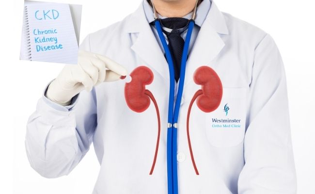
The kidneys are two bean-shaped organs. Each kidney is about the size of a fist. Your kidneys filter extra water and wastes out of your blood and make urine. Kidney disease means your kidneys are damaged and can’t filter blood the way they should.

Kidney Functions

  • Help remove waste and excess fluid
  • Filter the blood, keeping some compounds while removing others
  • Control the production of red blood cells
  • Make vitamins that control growth
  • Release hormones that help regulate blood pressure
  • Help regulate blood pressure, red blood cells, and the amount of certain nutrients in the body, such as calcium and potassium.

Chronic kidney disease includes conditions that damage your kidneys and decrease their ability to keep you healthy by doing the jobs listed above. If kidney disease gets worse, wastes can build to high levels in your blood and make you feel sick. You may develop complications like high blood pressure, anemia (low blood count), weak bones, poor nutritional health and nerve damage. Also, kidney disease increases your risk of having heart and blood vessel disease. These problems may happen slowly over a long period of time. Chronic kidney disease may be caused by diabetes, high blood pressure and other disorders. Early detection and treatment can often keep chronic kidney disease from getting worse. When kidney disease progresses, it may eventually lead to kidney failure, which requires dialysis or a kidney transplant to maintain life.

What causes CKD?

The two main causes of chronic kidney disease are diabetes and high blood pressure, which are responsible for up to two-thirds of the cases. Diabetes happens when your blood sugar is too high, causing damage to many organs in your body, including the kidneys and heart, as well as blood vessels, nerves and eyes. High blood pressure, or hypertension, occurs when the pressure of your blood against the walls of your blood vessels increases. If uncontrolled, or poorly controlled, high blood pressure can be a leading cause of heart attacks, strokes and chronic kidney disease. Also, chronic kidney disease can cause high blood pressure.

Other conditions that affect the kidneys are:

  • Glomerulonephritis, a group of diseases that cause inflammation and damage to the kidney’s filtering units. These disorders are the third most common type of kidney disease.
  • Inherited diseases, such as polycystic kidney disease, which causes large cysts to form in the kidneys and damage the surrounding tissue.
  • Malformations that occur as a baby develops in its mother’s womb. For example, a narrowing may occur that prevents normal outflow of urine and causes urine to flow back up to the kidney. This causes infections and may damage the kidneys.
  • Lupus and other diseases that affect the body’s immune system.
  • Obstructions caused by problems like kidney stones, tumors or an enlarged prostate gland in men.
  • Repeated urinary infections.

What are the symptoms of CKD?

Most people may not have any severe symptoms until their kidney disease is advanced. However, you may notice that you:

  • Feel more tired and have less energy
  • Have trouble concentrating
  • Have a poor appetite
  • Have trouble sleeping
  • Have muscle cramping at night
  • Have swollen feet and ankles
  • Have puffiness around your eyes, especially in the morning
  • Have dry, itchy skin
  • Need to urinate more often, especially at night.

Anyone can get chronic kidney disease at any age. However, some people are more likely than others to develop kidney disease. You may have an increased risk for kidney disease if you:

  • Have diabetes
  • Have high blood pressure
  • Have a family history of kidney failure
  • Are older
  • Belong to a population group that has a high rate of diabetes or high blood pressure, such as African Americans, Hispanic Americans, Asian, Pacific Islanders, and American Indians.

Diagnosing CKD

To check for kidney disease, health care providers use:

  • A blood test that checks how well your kidneys are filtering your blood, called GFR. GFR stands for glomerular filtration rate.
  • A urine test to check for albumin. Albumin is a protein that can pass into the urine when the kidneys are damaged.

If you have kidney disease, your health care provider will use the same two tests to help monitor your kidney disease and make sure your treatment plan is working.

What happens if my test results show I may have chronic kidney disease?

Your doctor will want to pinpoint your diagnosis and check your kidney function to help plan your treatment. The doctor may do the following:

  • Calculate your Glomerular Filtration Rate (GFR), which is the best way to tell how much kidney function you have. You do not need to have another test to know your GFR. Your doctor can calculate it from your blood creatinine, your age, race, gender and other factors. Your GFR tells your doctor your stage of kidney disease and helps the doctor plan your treatment.
  • Perform an ultrasound or CT scan to get a picture of your kidneys and urinary tract. This tells your doctor whether your kidneys are too large or too small, whether you have a problem like a kidney stone or tumor and whether there are any problems in the structure of your kidneys and urinary tract.
  • Perform a kidney biopsy, which is done in some cases to check for a specific type of kidney disease, see how much kidney damage has occurred and help plan treatment. To do a biopsy, the doctor removes small pieces of kidney tissue and looks at them under a microscope.

Your doctor may also ask you to see a Kidney specialist who will consult on your case and help manage your care.

Managing Chronic Kidney Disease

If you have chronic kidney disease (CKD), you can take steps to protect your kidneys from more damage.

The sooner you know you have kidney disease, the better. The steps you take to protect your kidneys from damage also may help prevent heart disease—and improve your health overall. Making these changes when you have no symptoms may be hard, but it’s worthwhile.

Ten ways to manage kidney disease

  1. Control your blood pressure
  2. Meet your blood glucose goal if you have diabetes
  3. Work with your health care team to monitor your kidney health
  4. Take medicines as prescribed
  5. Work with a dietitian to develop a meal plan
  6. Make physical activity part of your routine
  7. Aim for a healthy weight
  8. Get enough sleep
  9. Stop smoking
  10. Find healthy ways to cope with stress and depression

Eating Right for Chronic Kidney Disease

You may need to change what you eat to manage your chronic kidney disease (CKD). Work with a registered dietitian to develop a meal plan that includes foods that you enjoy eating while maintaining your kidney health.

The steps below will help you eat right as you manage your kidney disease. The first three steps (1-3) are important for all people with kidney disease. The last two steps (4-5) may become important as your kidney function goes down.

The first steps to eating right

Step 1: Choose and prepare foods with less salt and sodium

Why? To help control your blood pressure. Your diet should contain less than 2,300 milligrams of sodium each day.

  • Buy fresh food often. Sodium (a part of salt) is added to many prepared or packaged foods you buy at the supermarket or at restaurants.
  • Cook foods from scratch instead of eating prepared foods, “fast” foods, frozen dinners, and canned foods that are higher in sodium. When you prepare your own food, you control what goes into it.
  • Use spices, herbs, and sodium-free seasonings in place of salt.
  • Check for sodium on the Nutrition Facts label of food packages. A Daily Value of 20 percent or more means the food is high in sodium.
  • Try lower-sodium versions of frozen dinners and other convenience foods.
  • Rinse canned vegetables, beans, meats, and fish with water before eating.

Look for food labels with words like sodium free or salt free; or low, reduced, or no salt or sodium; or unsalted or lightly salted.

Step 2: Eat the right amount and the right types of protein

Why? To help protect your kidneys. When your body uses protein, it produces waste. Your kidneys remove this waste. Eating more protein than you need may make your kidneys work harder.

  • Eat small portions of protein foods.
  • Protein is found in foods from plants and animals. Most people eat both types of protein. Talk to your dietitian about how to choose the right combination of protein foods for you.

Animal-protein foods:

  • Chicken
  • Fish
  • Meat
  • Eggs
  • Dairy

A cooked portion of chicken, fish, or meat is about 2 to 3 ounces or about the size of a deck of cards. A portion of dairy foods is ½ cup of milk or yogurt, or one slice of cheese.

Plant-protein foods:

  • Beans
  • Nuts
  • Grains

A portion of cooked beans is about ½ cup, and a portion of nuts is ¼ cup. A portion of bread is a single slice, and a portion of cooked rice or cooked noodles is ½ cup.

Step 3: Choose foods that are healthy for your heart

Why? To help keep fat from building up in your blood vessels, heart, and kidneys.

  • Grill, broil, bake, roast, or stir-fry foods, instead of deep frying.
  • Cook with nonstick cooking spray or a small amount of olive oil instead of butter.
  • Trim fat from meat and remove skin from poultry before eating.
  • Try to limit saturated and trans fats. Read the food label.

Heart-healthy foods:

  • Lean cuts of meat, such as loin or round
  • Poultry without the skin
  • Fish
  • Beans
  • Vegetables
  • Fruits
  • Low-fat or fat-free milk, yogurt, and cheese

Step 4: Choose foods and drinks with less phosphorus

Why? To help protect your bones and blood vessels. When you have CKD, phosphorus can build up in your blood. Too much phosphorus in your blood pulls calcium from your bones, making your bones thin, weak, and more likely to break. High levels of phosphorus in your blood can also cause itchy skin, and bone and joint pain.

  • Many packaged foods have added phosphorus. Look for phosphorus—or for words with “PHOS”—on ingredient labels.
  • Deli meats and some fresh meat and poultry can have added phosphorus. Ask the butcher to help you pick fresh meats without added phosphorus.

Step 5: Choose foods with the right amount of potassium

Why? To help your nerves and muscles work the right way. Problems can occur when blood potassium levels are too high or too low. Damaged kidneys allow potassium to build up in your blood, which can cause serious heart problems. Your food and drink choices can help you lower your potassium level, if needed.

  • Salt substitutes can be very high in potassium. Read the ingredient label. Check with your provider about using salt substitutes.
  • Drain canned fruits and vegetables before eating.

Preventing Chronic Kidney Disease

You can protect your kidneys by preventing or managing health conditions that cause kidney damage, such as diabetes and high blood pressure. The steps described below may help keep your whole body healthy, including your kidneys.

1. Make healthy food choices

Choose foods that are healthy for your heart and your entire body: fresh fruits, fresh or frozen vegetables, whole grains, and low-fat or fat-free dairy products. Eat healthy meals, and cut back on salt and added sugars. Aim for less than 2,300 milligrams of sodium each day. Try to have less than 10 percent of your daily calories come from added sugars.

Tips for making healthy food choices

  • Cook with a mix of spices instead of salt.
  • Choose veggie toppings such as spinach, broccoli, and peppers for your pizza.
  • Try baking or broiling meat, chicken, and fish instead of frying.
  • Serve foods without gravy or added fats.
  • Try to choose foods with little or no added sugar.
  • Gradually work your way down from whole milk to 2 percent milk until you’re drinking and cooking with fat-free (skim) or low-fat milk and milk products.
  • Eat foods made from whole grains—such as whole wheat, brown rice, oats, and whole-grain corn—every day. Use whole-grain bread for toast and sandwiches; substitute brown rice for white rice for home-cooked meals and when dining out.
  • Read food labels. Choose foods low in saturated fats, trans fats, cholesterol, salt (sodium), and added sugars.
  • Slow down at snack time. Eating a bag of low-fat popcorn takes longer than eating a slice of cake. Peel and eat an orange instead of drinking orange juice.
  • Try keeping a written record of what you eat for a week. It can help you see when you tend to overeat or eat foods high in fat or calories.

2. Make physical activity part of your routine

Be active for 30 minutes or more on most days. If you are not active now, ask your health care provider about the types and amounts of physical activity that are right for you. Add more activity to your life with these tips to help you get active.

3. Aim for a healthy weight

If you are overweight or have obesity, work with your health care provider or dietitian to create a realistic weight-loss plan. View more weight control and physical activity resources to help you get and stay motivated.

4. Get enough sleep

Aim for 7 to 8 hours of sleep each night. If you have trouble sleeping, take steps to improve your sleep habits 

5. Stop smoking

If you smoke or use other tobacco products, stop. Ask for help so you don’t have to do it alone.

6. Limit alcohol intake 

Drinking too much alcohol can increase your blood pressure and add extra calories, which can lead to weight gain. If you drink alcohol , limit yourself to one drink per day if you are a woman and two drinks per day if you are a man. One drink is:

  • 12 ounces of beer
  • 5 ounces of wine
  • 1.5 ounces of liquor

7. Explore stress-reducing activities

Learning how to manage stress , relax, and cope with problems can improve emotional and physical health. Physical activity can help reduce stress, as can mind and body practices such as meditation, yoga , or tai chi.

8. Manage diabetes, high blood pressure, and heart disease

If you have diabetes, high blood pressure, or heart disease, the best way to protect your kidneys from damage is to

Keep blood glucose numbers close to your goal. Checking your blood glucose, or blood sugar, level is an important way to manage your diabetes. Your health care team may want you to test your blood glucose one or more times a day.

Keep your blood pressure numbers close to your goal. The blood pressure goal for most people with diabetes is below 140/90 mm Hg. Take all your medicines as prescribed. To help prevent heart attacks and stroke, keep your cholesterol levels in the target range. There are two kinds of cholesterol in your blood: LDL and HDL. LDL or “bad” cholesterol can build up and clog your blood vessels, which can cause a heart attack or stroke. HDL or “good” cholesterol helps remove the “bad” cholesterol from your blood vessels. A cholesterol test also may measure another type of blood fat called triglycerides.

Treatment of CKD

Some people live with kidney disease for years and are able to maintain kidney function. Others progress quickly to kidney failure.

Kidney failure means that your kidneys have lost most of their ability to function—less than 15 percent of normal kidney function. If your kidney function drops to this level, you may have symptoms from the buildup of waste products and extra water in your body.

To replace your lost kidney function, you may have one of three treatment options:

End-stage renal disease (ESRD) is kidney failure that is treated by dialysis or kidney transplant. Some people with kidney failure choose not to have dialysis or a transplant but continue to receive care from their health care team, take medicines, and monitor their diet and lifestyle choices.

Work with your health care professional, Dubai and family to consider your options and choose a treatment that is right for you.

Reference:

Disclaimer: All contents on this site are for general information and in no circumstances information be substituted for professional advice from the relevant healthcare professional, Writer does not take responsibility of any damage done by the misuse or use of the information.

February 28, 2021

Environmental allergies are an immune response to something in your surroundings that’s typically otherwise harmless. Symptoms of environmental allergies vary from person to person but can include sneezing, coughing, and fatigue. environmental allergies are a response to triggers you come into contact with in your surroundings or inhale during your day-to-day activities.

Symptoms

The symptoms of environmental allergies can be similar to a cold, but they’re not caused by the same thing. A virus causes a cold while allergies are a reaction caused by an immune system response to certain substances in your surroundings.

Some of the symptoms of environmental allergies include:

  • Sneezing
  • Runny nose
  • Shortness of breath
  • Itching
  • Headaches
  • Wheezing
  • Fatigue

Common environmental allergens

An allergen is anything that causes your immune system to trigger an allergic reaction. Identifying your allergens is an important first step in coming up with a treatment plan. These five environmental allergens are the most common.

Dust mites

Dust mites are one of the most common indoor allergens. They’re microscopic bugs that commonly live in furniture and mattresses in your home. If you have a dust mite allergy, your symptoms might be worse in the spring and summer months. That’s because dust mites prefer warm and humid environments.

Pollen

Pollen is another common allergen. If you’re allergic to pollen, your symptoms of sneezing, watery eyes, or an itchy throat might be worse when pollen increases in the spring and late fall.

Pet dander

Pet dander and pet saliva are common allergens. The symptoms of pet allergies can include:

  • Sneezing
  • Itching
  • Coughing
  • Hives

You may experience these symptoms if you’re around an animal, or if you’re in a home or car where an animal’s been. You may even have symptoms if a person near you has dander on their clothing.

Mold

Mold spores can cause mild to severe allergic reactions if you have a mold allergy. Symptoms of mold allergy may include:

  • Coughing
  • Trouble breathing
  • Sneezing
  • itchy skin

Mold thrives in damp environments, so your symptoms may be worse during months with damp weather. Mold is also commonly found in basements and bathrooms.

Cigarette smoke

Cigarette smoke has been found to irritate and worsen allergy symptoms in many people. Avoid smoking and exposure to secondhand smoke, especially if you have allergies.

How are environmental allergies diagnosed?

If you think you have an environmental allergy, see an Allergist Dr. Shahid Abbas, DHCC, Dubai. He will ask you questions about your symptoms and your medical and family history. Using this information along with the results from allergy testing, Dr. Shahid Abbas can identify specific allergens that may be causing your symptoms.

Allergy tests may include:

  • Skin prick test
  • Blood test
  • Elimination diet, if your doctor suspects you have a food allergen

Allergy tests determine the specific allergens causing your symptoms. Once your doctor has identified your allergens, he can suggest medications and treatment options.

Treatment

After diagnosis, your doctor may recommend medication for treating your allergies. You may find relief using over-the-counter (OTC) medications, such as an antihistamine.

Some of these medications can cause drowsiness. Be sure to read warning labels and talk with your doctor about which Over the counter medication may be right for you. OTC antihistamines may be more appropriate for seasonal environmental allergies since you won’t have to take them long-term. If your allergies are severe, your doctor may recommend prescription medications.

You may be a candidate for allergen immunotherapy, also called allergy shots. Allergen immunotherapy involves multiple shots given over the course of a few years. Allergy shots can improve and reduce symptoms for an extended period of time.

Home remedies and prevention tips

Preventing exposure to allergens can be one of the most effective ways to managing your symptoms and creating an allergy-free home. You may also be able to manage or reduce your symptoms with home remedies.

1. Use an air filter

Indoor air filters can improve the quality of indoor air by trapping allergens and pollutants before they enter your home. A 2018 study noted improved indoor air quality after installation of an air purifier equipped with a high-efficiency particulate air (HEPA) filter.

People with dust mite allergies also noted an improvement in symptoms, which suggested the air filter improved quality of life over the course of the study.

2. Allergy-proof your bed

Allergen-proof pillowcases and mattress covers can also be used to prevent exposure to dust mites. A diligent cleaning routine involving weekly washing of sheets and vacuuming can reduce exposure to allergens as well.

Make sure you wash your bedding in hot water to remove all allergens. Vacuuming your mattress while your bedding’s in the wash can also help reduce dust mites and, if you have a pet, dander.

3. Close your windows

Keeping your windows closed can help reduce the number of environmental allergens in your home, especially on days with a high pollen count.

The one part of your house where you should regularly open the windows, if you have them, is in the bathroom. Open windows or turn on a bathroom vent following showers to help remove moisture and prevent mold from growing.

4. Keep pets out of the bedroom

If you have pets, keep them out of your bedroom. Since you spend an extended period of time in your bedroom sleeping, having less allergens there can reduce symptoms. It may also improve your sleep.

Also, bathe your pets regularly to reduce the amount of dander. If your pet goes outside, regular baths can also reduce the risk of them tracking in allergens like pollen.

5. Take a probiotic

Researchers believe, there may be an association between gut bacteria and allergies, including seasonal allergies. In one study, participants who took probiotics reported improved seasonal allergy symptoms compared with those who took a placebo. However, the improvements seen may have been specific to one set of probiotics instead of all probiotics.

6. Use saline

OTC saline nasal spray has recently been found to be an effective way to manage symptoms of dust mite allergies. One 2016 study noted a significant improvement of coughing caused by allergies in participants who used saline solution nasal spray for 30 days.

7. Add essential oils

Essential oils can be used to support conventional treatment. Lavender, tea tree, and eucalyptus oils are anti-inflammatories and may provide relief from congestion and itchy or swollen eyes.

Full-strength essential oils can cause irritation or adverse reactions, so be sure to dilute them with a carrier oil or use a diffuser. Keep in mind that the U.S. Food and Drug Administration doesn’t regulate the purity, quality, or packaging of essential oils. Be sure to use essential oils as directed and purchase oils only from trusted, reputable sources.

8. Practice good hygiene

Showering after being outdoors can help remove allergens from your body. You should also wash your clothing if you’ve been digging in the yard or raking leaves. That can help prevent tracking mold spores and pollen into your home.

The takeaway

Environmental allergies can cause mild to severe symptoms, including sneezing, headaches, fatigue, and coughing. Preventing exposure to environmental allergens is the best way to treat them. You may not be able to avoid the allergens that trigger your symptoms. Your healthcare provider Dr. Shahid Abbas , Consultant Allergy and Immunology, DHCC, Dubai can help you create a plan for treating your allergies through home remedies and medications.

Reference:

  • oxfordhealth.nhs.uk
  • health.harvard.edu – https://www.health.harvard.edu/staying-healthy/3-ways-to-manage-allergies
  • Mayoclinic.org

Disclaimer: All contents on this site are for general information and in no circumstances information be substituted for professional advice from the relevant healthcare professional, Writer does not take responsibility of any damage done by the misuse or use of the information.

February 27, 2021

Hemorrhoids are the pillow-like clusters of veins that lie just beneath the mucous membranes lining the lowest part of the rectum and the anus. The condition most of us call hemorrhoids (or piles) develops when those veins become swollen and distended, like varicose veins in the legs. .

Who is more likely to get hemorrhoids?

You are more likely to get hemorrhoids if you:

  • Strain during bowel movements
  • Sit on the toilet for long periods of time.
  • Have chronic constipation or diarrhea
  • Eat foods that are low in fiber
  • Are older than age 50.
  • Are pregnant.
  • Often lift heavy objects

Types of Hemorrhoids

Internal hemorrhoids. Internal hemorrhoids lie inside the anal canal, where they primarily cause the symptom of intermittent bleeding, usually with bowel movements, and sometimes mucous discharge. They are usually painless. Internal hemorrhoids also may protrude (prolapse) outside the anus, where they appear as small, grape-like masses. Usually, the prolapsed hemorrhoid can be pushed back into the anus with a fingertip.

External hemorrhoids. These lie just outside the anal opening, where they primarily cause symptoms of swelling or bothersome protrusions, and sometimes discomfort. Swelling and discomfort may occur only intermittently. External hemorrhoids may also cause difficulties keeping the anal area clean after bowel movements. External hemorrhoids sometimes develop a blood clot inside of them (“thrombosis”), often after a period of diarrhea or constipation. In that case, it produces a sudden firm and painful swelling or lump around the rim of the anus.

Hemorrhoids are a quite common health problem. Hemorrhoids are more likely to develop in individuals who eat insufficient dietary fiber and do not get enough exercise, which can lead to repeated episodes of constipation and straining to have bowel movements.

Symptoms of hemorrhoids

The symptoms of hemorrhoids depend on the type you have.

If you have external hemorrhoids, you may have:

  • Anal itching
  • One or more hard, tender lumps near your anus
  • Anal ache or pain, especially when sitting

Too much straining, rubbing, or cleaning around your anus may make your symptoms worse. For many people, the symptoms of external hemorrhoids go away within a few days.

If you have internal hemorrhoids, you may have:

  • Bleeding from your rectum––bright red blood on stool, on toilet paper, or in the toilet bowl after a bowel movement
  • A hemorrhoid that has fallen through your anal opening, called prolapse

Internal hemorrhoids that are not prolapsed most often are not painful. Prolapsed internal hemorrhoids may cause pain and discomfort.

Diagnosis of Hemorrhoids

Your doctor can often diagnose hemorrhoids based on your medical history and a physical exam. He or she can diagnose external hemorrhoids by checking the area around your anus. To diagnose internal hemorrhoids, your doctor will perform a digital rectal exam and may perform procedures to look inside your anus and rectum.

Your doctor will ask you to provide your medical history and describe your symptoms. He or she will ask you about your eating habits, toilet habits, enema and laxative use, and current medical conditions.

Physical exam

Your doctor will check the area around your anus for

  • lumps or swelling
  • Internal hemorrhoids that have fallen through your anal opening, called prolapse.
  • External hemorrhoids with a blood clot in a vein
  • Leakage of stool or mucus
  • Skin irritation
  • Skin tags–extra skin that is left behind when a blood clot in an external hemorrhoid dissolve.
  • Anal fissures—a small tear in the anus that may cause itching, pain, or bleeding.

Your doctor will perform a digital rectal exam to

  • Check the tone of the muscles in your anus
  • Check for tenderness, blood, internal hemorrhoids, and lumps or masses

Treatment of Hemorrhoids

You can most often treat your hemorrhoids at home by

  • Eating foods that are high in fiber
  • Take warm sitz baths, especially when the hemorrhoids are uncomfortable. Sit in a tub or pan of plain warm water, 3 or 4 times a day, for 15-20 minutes each time. (Large pharmacies and medical supply stores also sell convenient plastic sitz bath devices that fit into a toilet). The water will keep the area clean, and the warmth will reduce inflammation and discomfort. Be certain to dry the rectal area thoroughly after each sitz bath. If you work, you can still take a sitz bath in the morning, upon returning from work, and again at bedtime.
  • Taking a stool softener or a fiber supplement
  • Drinking water or other nonalcoholic liquids each day as recommended by your health care professional.
  • Not straining during bowel movements
  • Not sitting on the toilet for long periods of time
  • Taking over-the-counter pain relievers
  • Sitting in a tub of warm water, called a sitz bath, several times a day to help relieve pain.

Applying over-the-counter hemorrhoid creams or ointments or using suppositories – a medicine you insert into your rectum may relieve mild pain, swelling, and itching of external hemorrhoids. Most often, doctors recommend using over-the-counter products for 1 week. You should follow up with your doctor if the products.

  • Do not relieve your symptoms after 1 week.
  • cause side effects such dry skin around your anus or a rash

Prevention

You can often prevent hemorrhoids by preventing constipation. Some of the following diet and lifestyle changes may help you to soften your stool, establish a regular schedule for bowel movements, and avoid the straining that can lead to hemorrhoids:

  • Add more fiber to your diet. Set a goal of 25 to 30 grams of fiber daily, from such high fiber foods as beans, broccoli, carrots, bran, whole grains and fresh fruits. Alternatively, many people find that it is more convenient to take a fiber powder such as psyllium (“Metamucil”), or methylcellulose (“Citrucel”), which is available at drug stores without prescription. To avoid bloating and gas, add fiber to your diet gradually over a period of several days.
  • Drink adequate amounts of fluid. For most healthy adults, this is the equivalent of 6 to 8 glasses of water daily.
  • Begin a program of regular exercise. As little as 20 minutes of brisk walking daily can stimulate your bowel to move regularly.
  • Train your digestive tract to have regular bowel movements. Schedule a time to sit on the toilet at approximately the same time each day. The best time to do this is usually right after a meal. Do not sit on the toilet for long periods (it tends to make hemorrhoids swell up and push out).
  • Respond immediately to the urge to have a bowel movement. Do not postpone until the time is more convenient.

Seek care right away!

You should seek medical care right away if you have severe anal pain and bleeding from your rectum, particularly with discomfort or pain in your abdomen, diarrhea, or fever. Also, see your doctor if you have severe rectal pain.

Eating, Diet, & Nutrition for Hemorrhoids

Your doctor may recommend that you eat more foods that are high in fiber. Eating foods that are high in fiber can make stools softer and easier to pass and can help treat and prevent hemorrhoids. Drinking water and other liquids, such as fruit juices and clear soups, can help the fiber in your diet work better. Ask your doctor about how much you should drink each day based on your health and activity level and where you live.

Prognosis

Hemorrhoids are not dangerous, and only need to be treated if they cause very bothersome symptoms. If hemorrhoids occur during pregnancy, they will usually regress spontaneously after childbirth. For hemorrhoids related to constipation, the prognosis is also good, provided you make the necessary changes to your diet and lifestyle. For hemorrhoids that cause persistent symptoms despite nonsurgical treatment, the results from office treatment or surgery are usually very good.

Reference

1. American Society of Colon and Rectal Surgeons
http://www.fascrs.org

2. National Institute of Diabetes & Digestive & Kidney Disorders
http://www.niddk.nih.gov/

3.  National Institute of Diabetes and Digestive and Kidney Diseases (NIDDK)

4. American Gastroenterological Association
http://www.gastro.org/

Disclaimer: All contents on this site are for general information and in no circumstances information be substituted for professional advice from the relevant healthcare professional, Writer does not take responsibility of any damage done by the misuse or use of the information.

Call Now Button